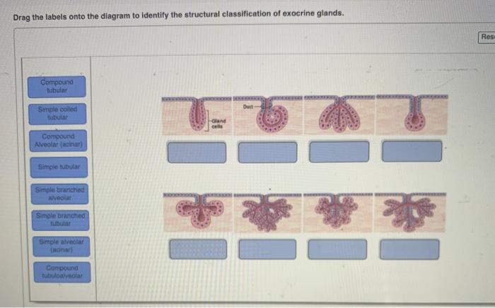
37 drag the labels onto the diagram to identify the structural classification of exocrine glands.
Ch 5-7 Lab A&P Mastering Flashcards - Quizlet Drag the labels onto the diagram to identify the basic structures of the epidermis-dermis junction. look at pic Drag the labels onto the diagram to identify the melanocyte in the stratum basale of the epidermis. Anatomy & Physiology II: Endocrine System - Quizlet Which endocrine structure synthesizes hormones involved with fluid balance and smooth muscle contraction? ... Drag the labels onto the diagram to identify the structural components of the hypophyseal portal system. ... The parathyroid Glands. Which is an effect of parathyroid hormone on peripheral tissues? Mobilizes calcium from bone.
Solved Drag the labels onto the diagram to identify the ... Anatomy and Physiology. Anatomy and Physiology questions and answers. Drag the labels onto the diagram to identify the structural classification of exocrine glands. Res Compound Dubular Simple coiled Oland Compound Alveolar (acinar) Simple tubular Simple branched hiver Simple branched Simple alveo (honan Compound.
Drag the labels onto the diagram to identify the structural classification of exocrine glands.
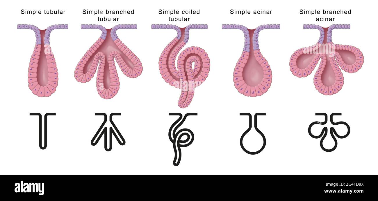
Exocrine Glands- Structure, Types And Examples Structure Of The Exocrine Glands. The structure of the exocrine glands is divided into two parts: Ductal portion. Glandular portion. The ductal portion is tubular in shape. It is a single, thick, cuboidal cell wall that helps in the movement of the secretion. The duct may be branched or unbranched. It can also be found as a simple coiled structure. EOF Solved Drag the labels onto the diagram to identify the ... Question: Drag the labels onto the diagram to identify the structural classification of exocrine glands. Reset Help Simple alveolar (acinar Duet Simple tubular Oland Simple branched tubular Simple colled Compound tubular . This problem has been solved!
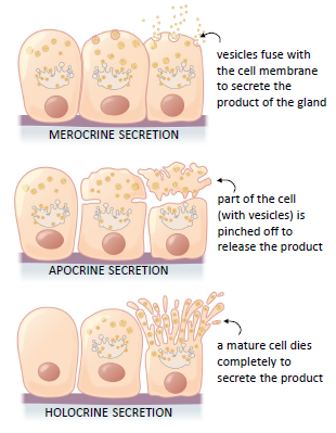
Drag the labels onto the diagram to identify the structural classification of exocrine glands.. HW 2.pdf - HW 2 Due: 11:59pm on Friday ... - Course Hero Correct Art-labeling Activity: A Structural Classification of Exocrine Glands Identify the structural classification of exocrine glands. Part A Drag the labels onto the diagram to identify the structural classification of exocrine glands. ANSWER: Help Reset Apocrine secretion Merocrine secretion Sebaceous gland Holocrine secretion Salivary ... Drag the labels onto the diagram to identify the - Course Hero Drag the labels onto the diagram to identify the different intercellular junctions found in animal tissues. ANSWER: Hint 3. How do substances move betw een plant cells? It is well known that cell walls in plants protect the cells from excessive uptake of water, but these walls also serve as a barrier to viruses. Solved Drag the labels onto the diagram to identify the ... Question: Drag the labels onto the diagram to identify the structural classification of exocrine glands. Reset Help Simple alveolar (acinar Duet Simple tubular Oland Simple branched tubular Simple colled Compound tubular . This problem has been solved! EOF
Exocrine Glands- Structure, Types And Examples Structure Of The Exocrine Glands. The structure of the exocrine glands is divided into two parts: Ductal portion. Glandular portion. The ductal portion is tubular in shape. It is a single, thick, cuboidal cell wall that helps in the movement of the secretion. The duct may be branched or unbranched. It can also be found as a simple coiled structure.
Neural Control of Human Movement
How are exocrine glands classified? | Socratic

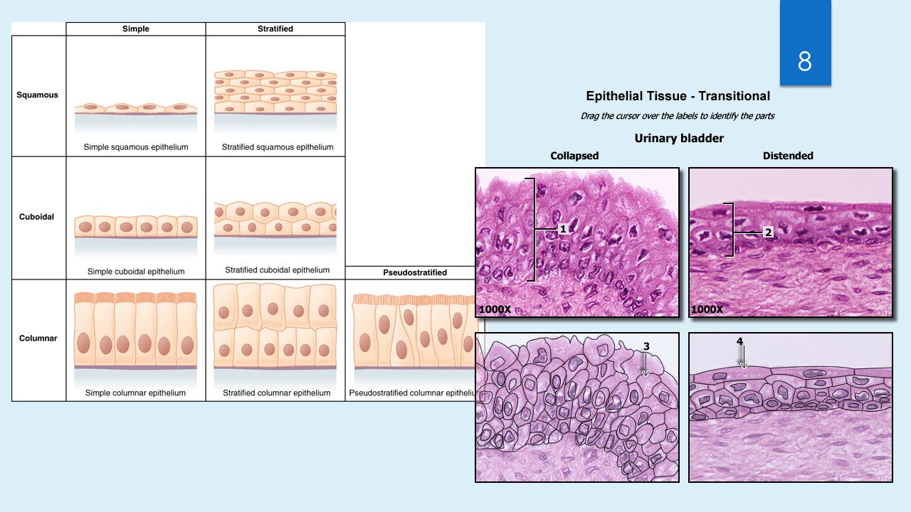
Epithelial Tissue | Anatomy and Physiology I

In vivo methods for drug absorption – Comparative ...
Training Manual on Primary Rehabilitation Therapy

EX 6 Review Sheet Flashcards | Quizlet
Glands: Anatomy and clinical notes | Kenhub

Lab Practice 4 -- Integumentary System Flashcards & Practice ...
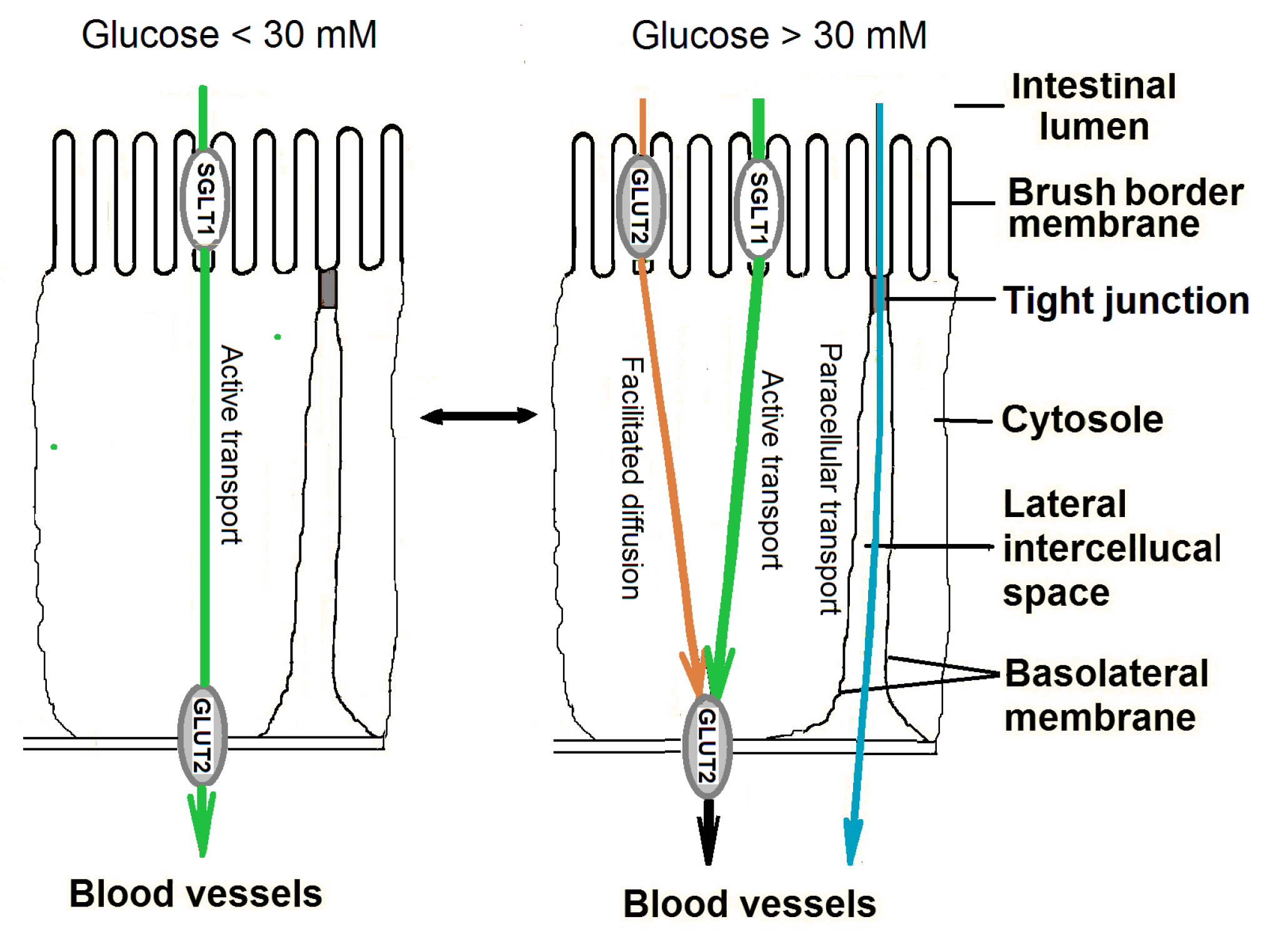
Nutrients | Free Full-Text | Mechanisms of Glucose Absorption ...

Ion Channels and Channelopathies
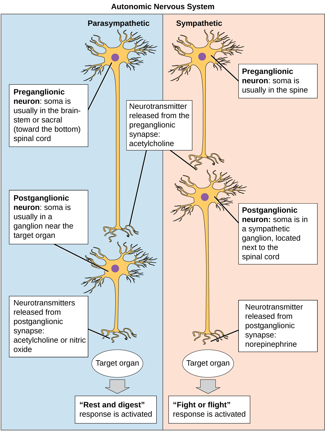
11.6 Nervous System – Concepts of Biology – 1st Canadian Edition

Gastric Acid, Calcium Absorption, and Their Impact on Bone ...
Making Meaning and Measurement in Gene Expression Analysis ...
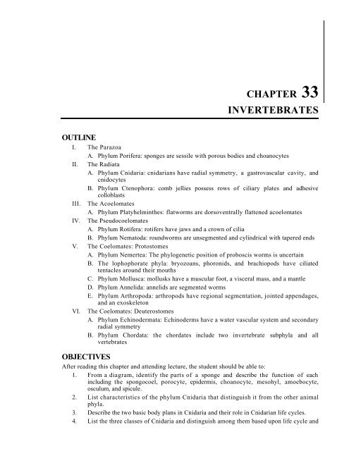
CHAPTER 33 INVERTEBRATES - BiologyJunction

Quiz #3 Study Guide Flashcards | Quizlet
Exocrine Gland High Resolution Stock Photography and Images ...
Building a Medical Terminology Foundation
Concepts of Biology

1.7 The Endocrine System – Neuroscience: Canadian 1st Edition ...

Epithelial Tissue | Anatomy and Physiology I
Untitled

PDF) Profiling the Triacylglyceride Contents in Bat ...

The Tissue Level of Organization - ppt download

1.7 The Endocrine System – Neuroscience: Canadian 1st Edition ...
Analyst

4.2 Epithelial Tissue – Anatomy & Physiology
Untitled
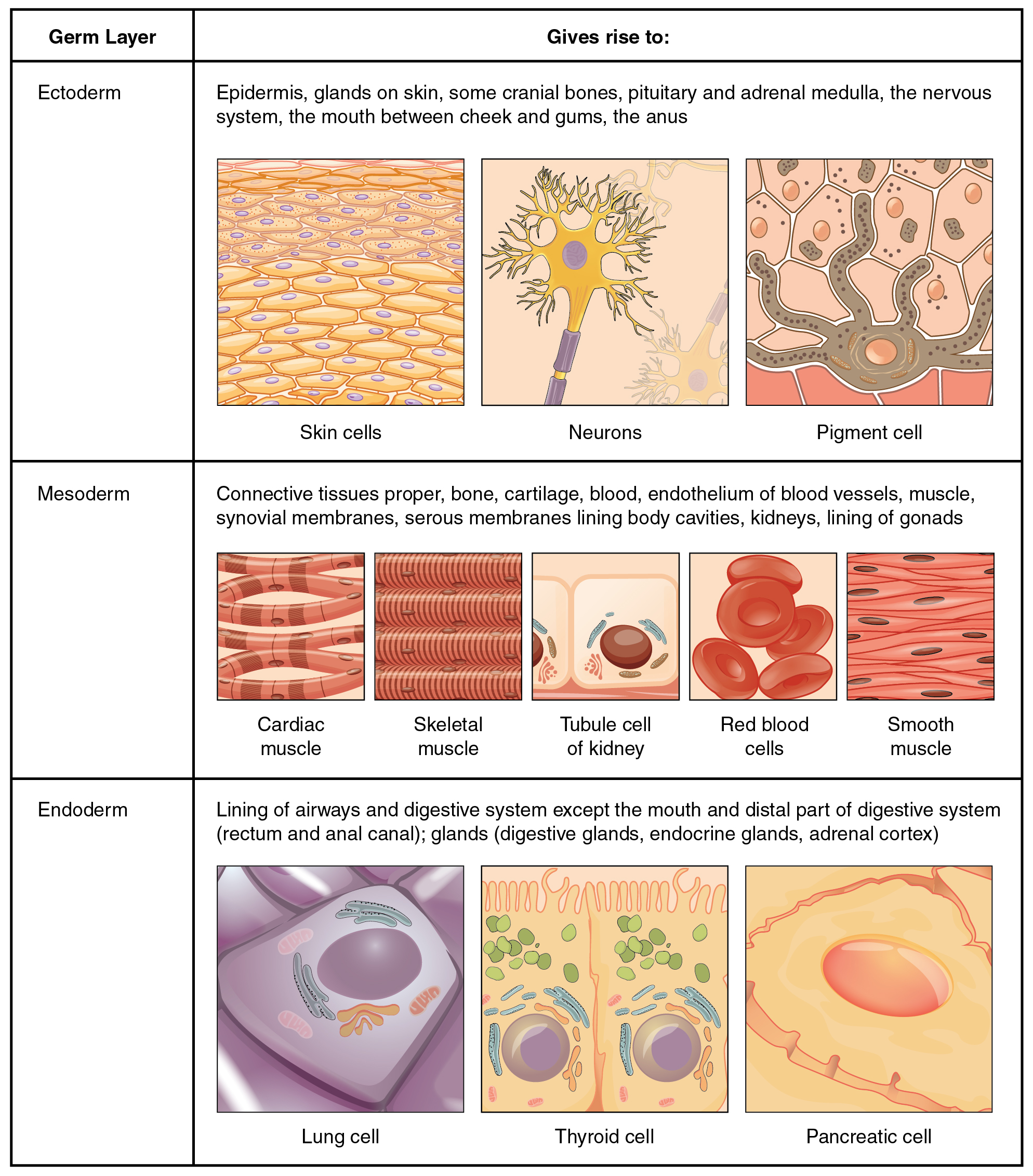
Types of Tissues – Anatomy & Physiology

HW 2.pdf - HW 2 Due: 11:59pm on Friday, September 22, 2017 To ...

HW 2.pdf - HW 2 Due: 11:59pm on Friday, September 22, 2017 To ...

1 1000 2782 http://uilis.unsyiah.ac.id/oer/files/original ...
SMART PHARMACOLOGY
Chapter 5 Key Terms 1 TendonOsteocytes ErythrocytesLigaments ...
Exocrine Gland High Resolution Stock Photography and Images ...

Learning objectives answered - Biol 121 - What is a cell ...

ALG: UGA Anatomy and Physiology 2 Lab Manual
Solved Drag the labels onto the diagram to identify the ...
0 Response to "37 drag the labels onto the diagram to identify the structural classification of exocrine glands."
Post a Comment