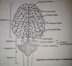
36 diagram of sheep brain
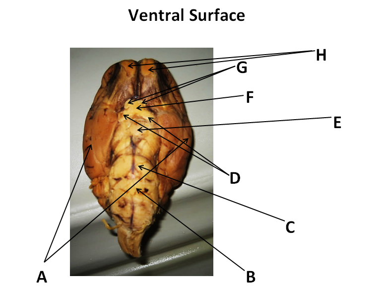
Exercise 19: Dissection of the Sheep Brain Flashcards - Easy Notecards In your own words, describe the firmness and texture of the sheep brain tissue as observed when cutting into IT. Felt tough, and rubbery. When cutting into, IT gets alot softer as you cut through. Diagram of Sheep Brain - Inferior view
Difference Between Human and Sheep Brain | Difference Between Human vs Sheep Brain There are a few differences between the human and sheep brain. The human brain is larger in size and shape when compared to the sheep's brain. Sheep brains do not have as many...

Diagram of sheep brain
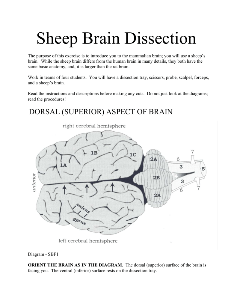
Sheep Brain Dissection - DocsBay Sheep brain dissection: pre-lab. (Pre-Lab must be submitted to start the lab). Part 1: Planes and Axis of the Brain. 1. Label the diagram to the right with the dorsal-ventral axis and the anterior-posterior axis. 2. Name the view of the brain shown in the diagrams below PDF sg_sheep_brain_dissection Sheep Brain Dissection Guide. Student Name: First Dissection -- Exploring the Neuron and its Parts. 1. Your teacher will draw a neuron on the board. 2. Place your sheep brain with the cut side (MEDIAL) down. Using the plastic knife, cut the brain about 1 inch from the rostral end (where the dent is located). Sheep Brain Dissection with Labeled Images The sheep brain is exposed and each of the structures are labeled and described in a sequential manner, in the same way that a real 1. The sheep brain is enclosed in a tough outer covering called the dura mater. You can still see some structures on the brain before you remove the dura mater.
Diagram of sheep brain. This online quiz is called Sheep Brain Diagram Sheep Brain Diagram. a quiz by epaz4689. • 18 plays. This is an online quiz called Sheep Brain Diagram. There is a printable worksheet available for download here so you can take the quiz with pen and paper. Sheep Brain Dissection by Lewis Heinzlmeir Sheep Brain Dissection. What is the significance of these convolutions? How do they provide mammals with an advantage? The convolutions are significant to mammals because it provides the brain with alot of surface area for brain cells and neuron pathways. List the cerebral lobes and describe the major... Frontiers | In vivo Diffusion Tensor Magnetic Resonance Tractography... Unraveling the principal brain texture features of preclinical models that are advantageously exploited in experimental neuroscience is crucial to correctly evaluate investigational findings and to correlate them with real clinical scenarios. Although structurally similar to the human brain, the gyrencephalic ovine... Sheep Brain Dissection Guide - Contents - StuDocu Sheep brains are used in this lab because they are easy to extract, reasonably inexpensive (they are procured from the food industry), large, and mammalian. A structure is anterior to another structure when it is closer to the nose of an animal (see the above diagram). Some texts use the terms anterior...
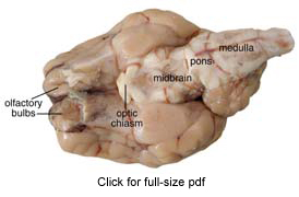
Sheep Brain Explora/on Guide - PDF Free Download Sheep Brain Explora/on Guide Sheep Brain Exploration Introduction: This guide is intended to lead you through the anatomy of the sheep brain dissection and also to make As illustrated in the human brain diagram below, there are two optic nerves that bring visual information from the eye to the brain. Sheep Brain: Cerebral Cortex, Cerebrum, Corpus Callosum, Olfactory... Human Brain Diagram. Cranial Nerves Coloring. Sheep Brain: Olfactory Tracts (CN I), Optic nerve (CN II), Olfactory Bulbs, Cerebellum, Pons, Medulla. Sheep brain dissection | Human Anatomy and Physiology Lab... The sheep brain is quite similar to the human brain except for proportion. Compare the sheep brain to the human brain. What do you notice about the size difference of each structure? Identify the clublike olfactory bulbs on the inferior surface of the frontal lobes of the cerebral hemispheres. PDF Dissection of the sheep's brain The purpose of the sheep brain dissection is to familiarize you with the three-dimensional structure of the brain and teach you one of the great methods While the course will emphasize the human brain, observation and evolution indicate that there are many similarities between the sheep brain and the...
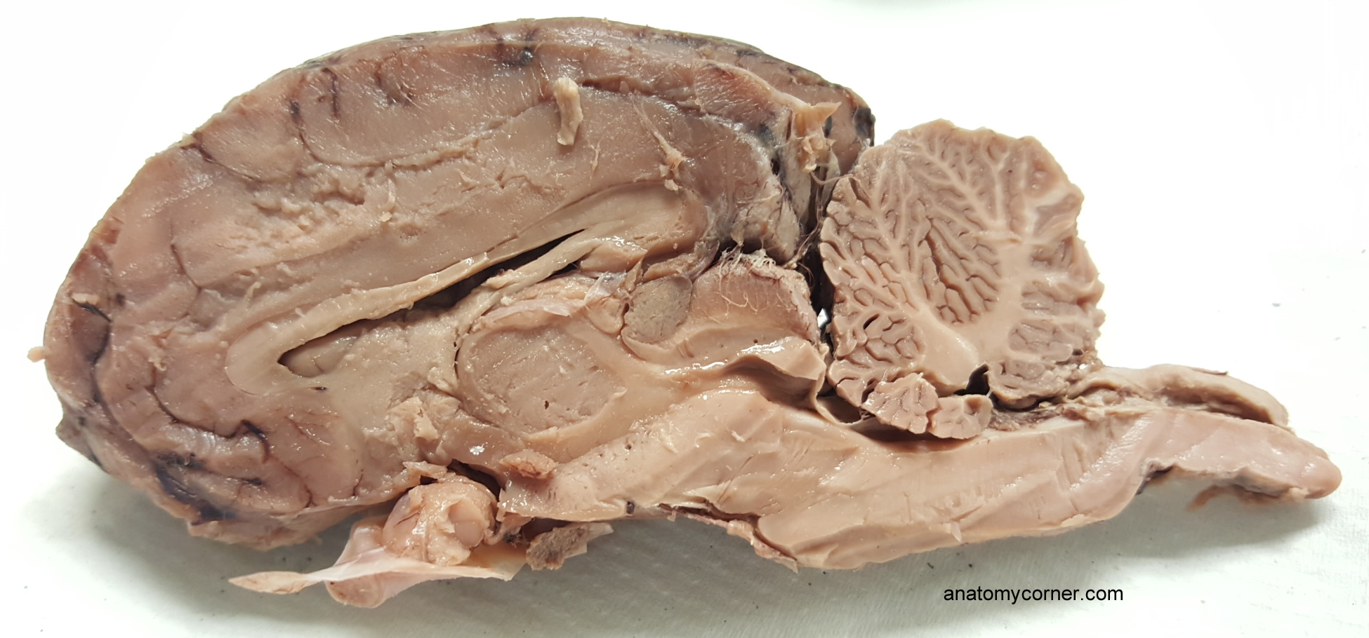
Sheep Brain Ventricles) Sheep Brain — Median View. Brainstem components of the ventricular system are visible in this median view of a sheep brain. The third ventricle is indicated by yellow pics. Sheep Brain Images Nervous system - sheep brain images. Sheep Brain Unlabeled. Sheep Brain Dissection Guide PowerPoint Presentation Sheep Brain Dissection Guide. Good Luck!!. Laurie Hayes. To begin. Get gloves and a rubber apron or lab coat. Sheep Brain Dissection Guide. Good Luck!!. Laurie Hayes. Label your diagram with parts of midbrain and brain stem • What is the function of the medulla oblongata and pons? • Sheep Brain vs Human Brain - Perception and Vision | Coursera Perception is how the brain interprets incoming stimuli. Not all stimuli that can be sensed are perceivable, and sometimes ... This is the human brain. Let's look at it from the side. Sheep, and human. Is it, is it going well Sol? >> Your glove is covering most of the sheep. >>
sheep brain midsagittal Diagram | Quizlet
7th Grade Science - Mrs. Donovan sheep brain dissection sheep brain dissection. Prerequisites: Module Completed Module In Progress Module Locked. Diagram of Sheep Brain Page.

Sheep Brain Images
Sheep brain - Big Chemical Encyclopedia Sheep brain. Provitamin D. Provitamin is made from cholesterol, and its commercial production begias with the Brain extracts firm BSE cattle have transmitted the disease to mice, sheep, cattle, pigs and monkeys. Studies of 12 recent cases of atypical CJD in the UK have provided evidence that the...

sheep brain pt 1 Diagram | Quizlet
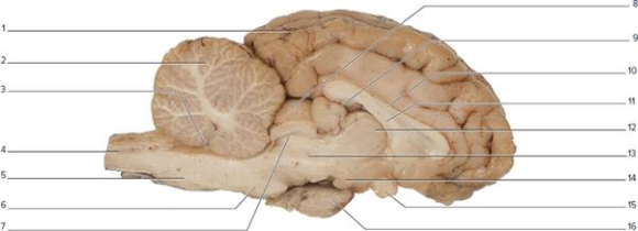
Lab Exam 3: Anatomy of Sheep Brain; Histology Flashcards - Cram.com Study Flashcards On Lab Exam 3: Anatomy of Sheep Brain; Histology at Cram.com. Quickly memorize the terms, phrases and much more. Diagram of sheep brain, dorsal view. Neuron cells (400x). Nerve and blood vessels slide (closeup of nerve).

Sheep Brain V (Lateral view) Diagram | Quizlet
A virtual sheep brain dissection guides anatomy studies with photos... The sheep brain is exposed and each of the structures are labeled and described in a sequential manner, in the same way that a real dissection Learn the external and internal anatomy of sheep brains with HST's Learning Center science lesson and guide! Diagram worksheets also included.
Lab 14 Sheepbraindiss 2
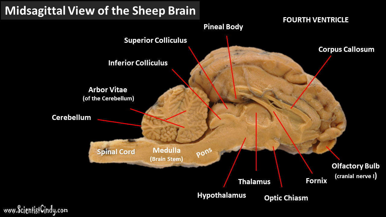
Neuron/Spinal Cord Histology Brain Anatomy Sheep Brain Dissection 26 Sheep Brain Photo from: 13 1 12 7 5 11 8 2 9 10 3 6 4 1. Corpus callosum 5. Pineal gland (body) Fourth ventricle Lateral ventricle 2. Thalamus 6. Mammillary Body Brain stem 3. Hypothalamus 7. Superior colliculus Arbor vitae (white matter) 4. Optic chiasm 8. Inferior colliculus Cerebellar gray...

Sheep Brain Images
PDF Sheep Brain Dissection Compare the size of the olfactory bulbs in a sheep relative to the total brain size compared to the Think about the different components of a neuron (see diagram below) and how this may influence Observe the sheep brain that has had the cerebellum and the caudal portion of the cerebral cortex...

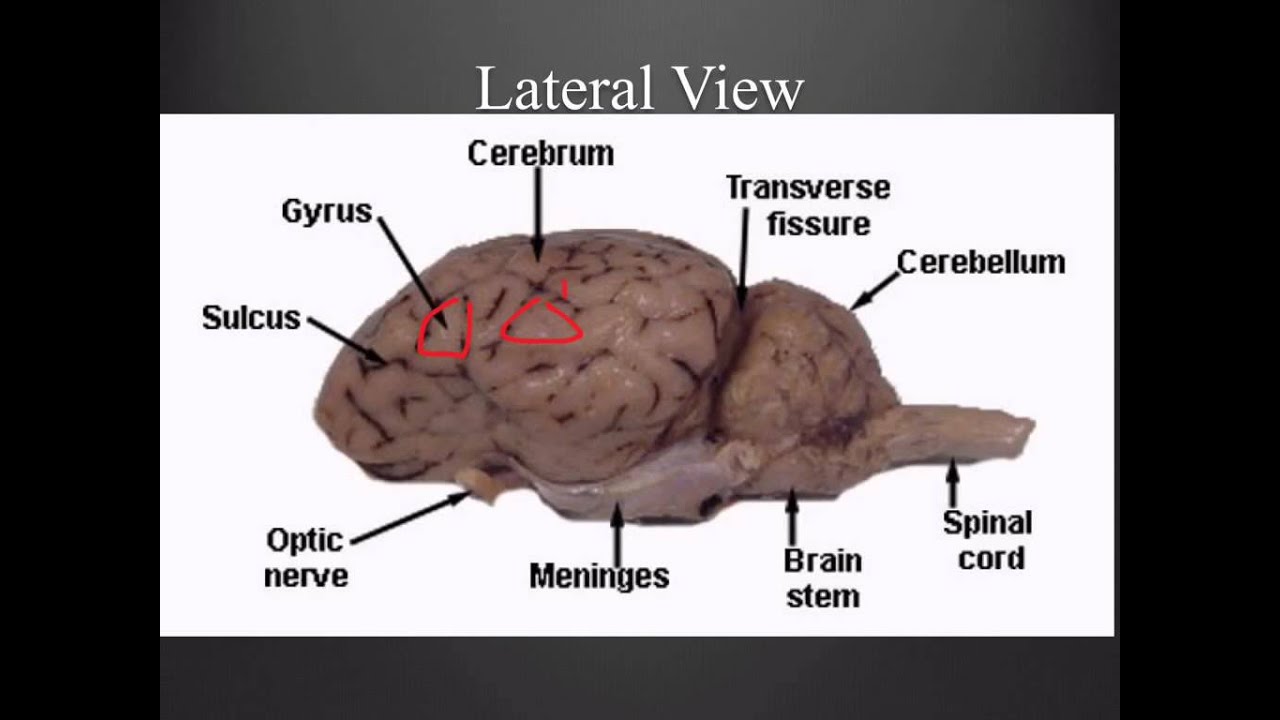
Cerebrum Sheep Dissection - Human Anatomy - GUWS Medical
Sheep Brain & Eye (with labels) - YouTube A video tutorial of the anatomy of the brain and eye of a sheep for comparative anatomy.
Lab Exam 3: Anatomy of Sheep Brain; Histology Flashcards ...
Sheep Brain Diagram - Free Catalogs A to Z Sheep Brain Dissection Project Guide - HST Learning Center. 8 hours ago Diagram Worksheets. Label the Parts of a Sheep Brain. Print out these diagrams and fill in the labels to test your knowledge of sheep brain anatomy. Internal anatomy: label the right side (.pdf) External anatomy: label the top...

Sheep Brain Dissection labeled Diagram | Quizlet
How Does a Sheep Brain and a Human Brain Compare? Sheep brains have less ridges and contours in comparison to human brains. While a human brain has more of a rounded shape, a sheep's brain is Sheep brains do not have these capabilities. Metacognition and other advanced cognitive skills, such as social intelligence, planning and reasoning...
Sheep Brain Dissection Picture Guide
Sheep Brain Dissection Guide Good Luck!! Ms. Lecce 8. Human vs Sheep • Compare the various areas of the sheep brain (cerebrum, brain stem, cerebellum) to the human brain. #4. How is it the same and How is it different? Label your diagram with parts of midbrain and brain stem • What is the function of the medulla oblongata and pons? •
Sheep Brain Dissection Picture Guide
DOC Sheep Brain Anatomy Lab Manual The brain of the sheep is useful for study because its anatomy is similar to human brain anatomy. Although exact proportions (and names) sometimes differ, every structure you will identify in the sheep brain corresponds to a homologous structure, usually with the same name, in humans.
Sheep Brain Dissection
Diagram of Sheep Brain | Quizlet Start studying Sheep Brain. Learn vocabulary, terms and more with flashcards, games and other study tools. the path along which the olfactory receptors send their electrical messages to the brain.

Sheep brain dissection | Human Anatomy and Physiology Lab ...
Sheep Brain Dissection with Labeled Images The sheep brain is exposed and each of the structures are labeled and described in a sequential manner, in the same way that a real 1. The sheep brain is enclosed in a tough outer covering called the dura mater. You can still see some structures on the brain before you remove the dura mater.
Sheep Brain DissectionLabel the following figure of a mids ...
PDF sg_sheep_brain_dissection Sheep Brain Dissection Guide. Student Name: First Dissection -- Exploring the Neuron and its Parts. 1. Your teacher will draw a neuron on the board. 2. Place your sheep brain with the cut side (MEDIAL) down. Using the plastic knife, cut the brain about 1 inch from the rostral end (where the dent is located).
Sheep Brain Explora on Guide
Sheep Brain Dissection - DocsBay Sheep brain dissection: pre-lab. (Pre-Lab must be submitted to start the lab). Part 1: Planes and Axis of the Brain. 1. Label the diagram to the right with the dorsal-ventral axis and the anterior-posterior axis. 2. Name the view of the brain shown in the diagrams below
Sheep Brain Dissection Guide
Sheep Brain Explora on Guide

Internal Brain Anatomy (sheep) Quiz
Topic: Sheep Brain Dissection Grades: 8-12th Number of ...
Sheep Brain Dissection

Sheep Brain Dissection | Carolina.com

sheep brain (sagittal) Diagram | Quizlet
Sheep Brain

Adventures with animals and plants. Biology. Fig. 255 A ...
The Brain - SCIENTIST CINDY

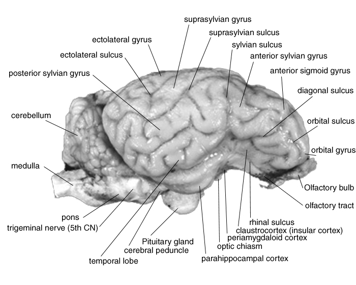
Identification of the motor areas in the sheep brain (left ...

Diagram of Sheep Brain - Inferior view

On the Cutting Edge: Exploring Sheep Organs | Carolina.com

Sheep Brain Dissection Guide - ppt download

Sheep Brain Diagram
Sheep Brain Dissection Project Guide | HST Learning Center
Sheep Brain Dissection Guide - ppt download

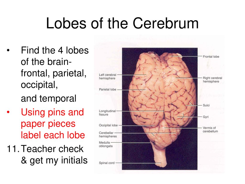
Lobes - sheep brain
The Brain Biodiversity Bank at Michigan State University
Sheep Brain Neuroanatomy Online Self-Test | KPU.ca - Kwantlen ...

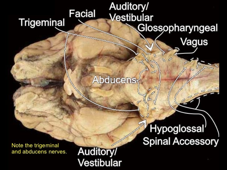
Sheep brain/Label Nerves Diagram | Quizlet
0 Response to "36 diagram of sheep brain"
Post a Comment