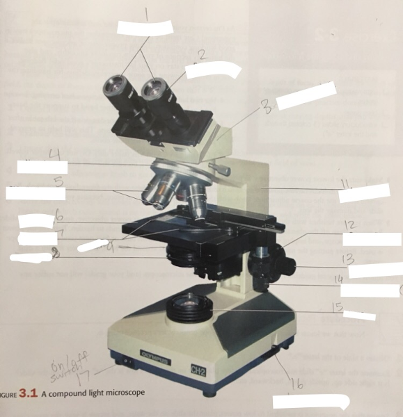
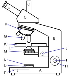
38 labeled diagram of a microscope
Microscope Parts And Functions Worksheet Answer Key Microscope Parts and Use Worksheet Answer Key or A Study Of the Microscope and Its Functions with A Labeled Diagram Microscope Parts and Use Worksheet Answer Key with A School Called Home A View Through the Microscope Identifying. In this article we have 20 great images on Microscope Parts And Use Worksheet Answer Key. 22 Parts Of a Microscope With Their Function And Labeled ... 22 Parts Of a Microscope With Their Function And Labeled Diagram. April 7, 2021 April 7, 2021 by Biocheminsider. Microscope Description. A microscope is a laboratory instrument used to examine objects that are too small to be seen by the naked eye. In other words, it enlarges images of small objects. ... Light microscope and Electron microscope ...
Jejunum Histology Slide with Labeled Diagram and ... Jejunum histology slide labeled diagram. Now, it is better to review all the histological features from the jejunum slide. I will try to show you all the features from the jejunum histology slide with the labeled diagram. Here, I tried to show you all the histological features from the four different layers of the jejunum wall.

Labeled diagram of a microscope
Compound Microscope- Definition, Labeled Diagram ... The optical microscope often referred to as the light microscope, is a type of microscope that uses visible light and a system of lenses to magnify images of small subjects. There are two basic types of optical microscopes: Simple microscopes. Compound microscopes. The term "compound" in compound microscopes refers to the microscope having ... Labeled Microscope Diagram - Tim's Printables 1 thought on "Labeled Microscope Diagram" Synovia A Jacobs. 06/28/2021 at 11:41 pm. Thank you these really helps. Reply. Leave a Reply Cancel reply. Your email address will not be published. Required fields are marked * Comment * Name* Email* Website. Light Microscope- Definition, Principle, Types, Parts ... A light microscope is a biology laboratory instrument or tool, that uses visible light to detect and magnify very small objects and enlarge them. They use lenses to focus light on the specimen, magnifying it thus producing an image. The specimen is normally placed close to the microscopic lens.
Labeled diagram of a microscope. Parts of the Microscope with Labeling (also Free Printouts ... Click to Download : Label the Parts of the Microscope with answers (A4) PDF print version. For a thorough review of each microscope part continue reading…. A basic microscope has a single convex lens such as those found in a magnifying glass, which you can use to visualize the finest prints. Labelled Diagram Of A Plant Cell Under A Microscope ... Animal Cell Diagram Electron Microscope. 11 is a labelled diagram of a leaf palisade mesophyll cell as seen With a high quality light microscope. But at the same time. Under the microscope Priya observes a cell that has a cell wall and a distinct nucleus. Its a thin slice. Parathyroid Gland Histology with Microscope Slide Image ... The sample tissue section and diagram also show the numerous fat cells (adipose tissue). You may join anatomy learner on social media for a more updated labeled diagram on the parathyroid gland. Parathyroid gland microscope slide image drawing. This is a straightforward task to draw the microscope slide image of the parathyroid gland. Plant Cell Under Light Microscope Labeled - Diagram Sketch Plant Cell Under Light Microscope Labeled. angelo on October 4, 2021. Editible Eps Vector File The Animal Cell Diagram Vector Etsy In 2021 Animal Cell Cell Diagram Plant Cell Diagram. Onion Epidermis Under Light Microscope Purple Colored Large Cells Project Microscopic Photography Epidermis.
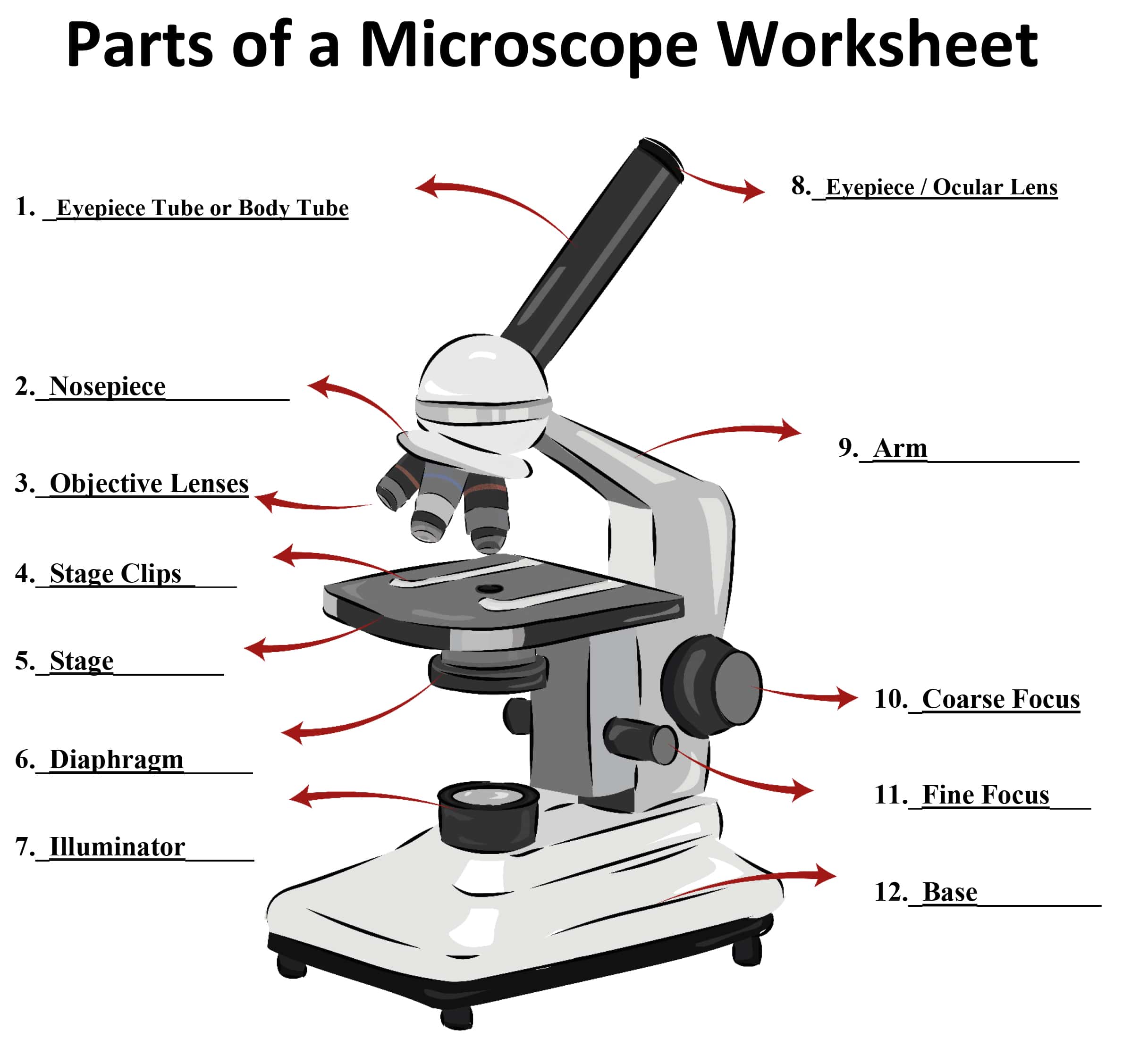
Microscope Diagram Worksheet - The Microscope Create A ... Write the letter on the line that represents each part of the microscope. Using the terms listed below, label the microscope diagram. When you can identify a part of the microscope place the . There is a printable worksheet available for download here so you can take the . Use the words from this word list to identify the parts of the microscope. Microscope, Microscope Parts, Labeled Diagram, and Functions Microscope, Microscope Parts, Labeled Diagram, and Functions Published by Admin on January 19, 2022 January 19, 2022. What is Microscope? A microscope is a laboratory instrument used to examine objects that are too small to be seen by the naked eye. It is derived from Ancient Greek words and composed of mikrós, "small" and skopeîn,"to ... Awesome Microscope Labeling Worksheet Answers - Labelco Labeling the parts of the microscope. Worksheet identifying the parts of the compound light microscope. Microscope parts and use worksheet answer key along with labeling the parts of the microscope blank diagram available for. Can be rotated to change magnification. Elodea Leaf Cell Labeled Diagram - Diagram Sketch Elodea Leaf Cell Labeled Diagram. angelo on February 20, 2022 Leave a Comment on Elodea Leaf Cell Labeled Diagram. ... Plant Cell Electron Microscope Worksheet Cell Diagram Plant Cell Plant Cell Diagram . Cell Transport Lab Osmosis And Diffusion Cell Transport Cell Osmosis .
Electron Microscope Principle, Uses, Types and Images ... Ans: A light microscope has a low resolving power (0.25µm to 0.3µm) while the electron microscope has a resolution power about 250 times higher than the light microscope at about 0.001µm. Similarly, a light microscope has a magnification of 500X to 1500x while the electron microscope has a much higher magnification of 100,000X to 300,000X. 39 microscope labeling worksheet answers - Worksheet Master Label Microscope Diagram - EnchantedLearning.com Answers Go to a microscope definition worksheet to print. EnchantedLearning.com Label Microscope Diagram. Using the terms listed below, label the microscope diagram. Inventions and Inventors. arm - this attaches the eyepiece and body tube to the base. base - this supports the... Fully Labeled Diagram Of Animal Cell - Studying Diagrams Plant Cell Diagram Electron Microscope The Greatest Garden In 2021 Plant Cell Diagram Cell Diagram Animal Cell Structure . Identify reactive astrocytes as critical mediators of vascular repair and remodeling after stroke. Fully labeled diagram of animal cell. This pdf packet contains 6 versions of the diagram to help you teach and also quiz ... Plant Cell And Animal Cell Labeled Diagram - Studying Diagrams Animal Cell- Definition Organelles Structure Parts Functions Labeled Diagram Worksheet Types of Plant Cell- Definition Structure Functions Labeled Diagram Amazing 27 things under the microscope diagrams and. 2 3 Eukaryotic Cells Bioninja Plant Cell Diagram Cell Diagram. Animal And Plant Cell Labeled.
Parts of Microscope (Labeling) Diagram | Quizlet
Microscope Types (with labeled diagrams) and Functions Phase-contrast microscope labeled diagram. Phase-contrast microscope functions: Its applications areas include. In cases where the specimen is colorless and is very tiny; In biology to conduct cellular level examination of microorganisms that can't be visualized using the bright field microscopy Interference Microscope

5 Important Types of Microscopes used in Biology (With Diagram)
Diagram Of Animal Cell As Seen Under Light Microscope ... Now the first thing to point out when looking at images under an electron microscope is the scale. The diagram is very clear and labeled. Its a thin slice. A one cell membrane B one cell wall C two cell membranes D two cell walls 3 The diagram shows a. So lets begin by drawing a rough-oval shape. Answer 1 of 2.
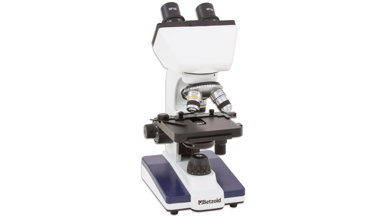
Betzold Binokulares Mikroskop Bin-TOP 02 mit LED-Auf- und Durchlicht
Compound Microscope - Diagram (Parts labelled), Principle ... Image : Labeled Diagram of compound microscope parts. See: Labeled Diagram showing differences between compound and simple microscope parts Structural Components. The three structural components include. 1. Head. This is the upper part of the microscope that houses the optical parts. 2. Arm . This part connects the head with the base and ...

Solved tration Questions: (10 points) Label the diagram of a ...
Labeled Microscope Worksheet Answers - Worksheet Academy Labeled microscope worksheet answers. High power objective 6. Students label the parts of the microscope in this photo of a basic laboratory light microscope. Each microscope layout both blank and the version with answers are available as pdf downloads. When focusing a specimen you should always start with the objective.

A labeled diagram of a microscope. MLT 101. :) | Teaching ...
All Microscope Parts | Function | Labeled Diagram ... Microscope parts labeled diagram gives us all the information about its parts and their position in the microscope. Microscope Parts Labeled Diagram The principle of the Microscope gives you an exact reason to use it. It works on the 3 principles. Magnification Resolving Power Numerical Aperture. Parts of Microscope Head Base Arm Eyepiece Lens

Compound Microscope Parts, Functions, and Labeled Diagram ...
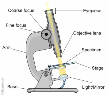
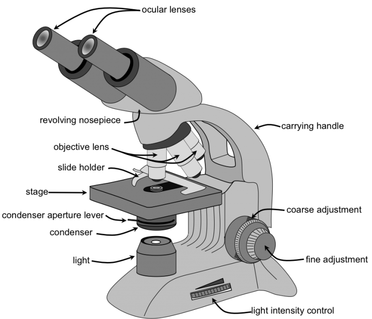
Parts of a microscope with functions and labeled diagram Figure: Diagram of parts of a microscope. There are three structural parts of the microscope i.e. head, base, and arm. Head - This is also known as the body, it carries the optical parts in the upper part of the microscope. Base - It acts as microscopes support. It also carries microscopic illuminators.
Guide to Using a Microscope - Year 8 Portfolio
Tonsil Histology Slide with Labeled Diagram - Histological ... The lighter area of all lymphatic nodules in the germinal center (shown in the diagram). Again, the labeled diagram also shows some diffuse lymphatic nodules present in the tonsil structure. A dense connective tissue capsule is also marked in the labeled diagram underlies the palatine tonsil.

Label The Microscope Parts! Diagram | Quizlet
Cecum Histology Slide with Labeled Image and Diagram ... Here, I will show you all the histological structures from the cecum with a microscope slide image and labeled diagram. I will also provide the appropriate identification points for the cecum slide under the light microscope. Again, you will get a little information on the specific histological features of the cecum in a different animal.

Draw a well labelled diagram of a microscope. - Brainly.in
Ruminant Reticulum Histology Slide with Labeled Diagram ... Again, the reticulum labeled diagram shows the inner oblique pattern of the smooth muscle layer. The outer smooth muscle layer of the labeled diagram shows the cross pattern. In addition, the tunica serosa layer of the reticulum microscope labeled image shows a thin layer of loose connective tissue with numerous blood vessels.
Microscope Parts and Function
Plant Cell Under Microscope Labeled - Diagram Sketch Plant Cell Under Microscope Labeled. Onion Epidermis Under Light Microscope Purple Colored Large Cells Project Microscopic Photography Epidermis. 40x 400x Compound Monocularbiological Microscope45 Degree Angled Headelectric Lightedbeginner Slides Plant Cell Things Under A Microscope Plant Cell Picture.

Parts of a compound microscope | Biology labs, Medical ...
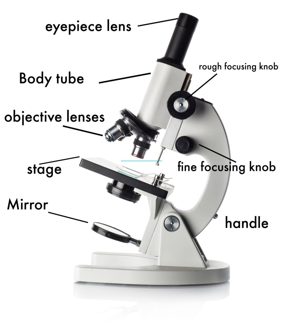
Simple Microscope - Diagram (Parts labelled), Principle ... Labeled Diagram of simple microscope parts Optical parts The optical parts of a simple microscope include Lens Mirror Eyepiece Lens A simple microscope uses biconvex lens to magnify the image of a specimen under focus.
Labelled Diagram of Microscope Parts
Light Microscope- Definition, Principle, Types, Parts ... A light microscope is a biology laboratory instrument or tool, that uses visible light to detect and magnify very small objects and enlarge them. They use lenses to focus light on the specimen, magnifying it thus producing an image. The specimen is normally placed close to the microscopic lens.

Label the Microscope Diagram | Download Scientific Diagram
Labeled Microscope Diagram - Tim's Printables 1 thought on "Labeled Microscope Diagram" Synovia A Jacobs. 06/28/2021 at 11:41 pm. Thank you these really helps. Reply. Leave a Reply Cancel reply. Your email address will not be published. Required fields are marked * Comment * Name* Email* Website.

How To Draw A Microscope, Step by Step, Drawing Guide, by ...
Compound Microscope- Definition, Labeled Diagram ... The optical microscope often referred to as the light microscope, is a type of microscope that uses visible light and a system of lenses to magnify images of small subjects. There are two basic types of optical microscopes: Simple microscopes. Compound microscopes. The term "compound" in compound microscopes refers to the microscope having ...
Lab Exam 1 - Labeling Diagrams Flashcards - Easy Notecards

All Microscope Parts | Function | Labeled Diagram | slidingmotion

BIOLOGY FROM 1 | EQUIPMENTS USED FOR OBSERVATION | Cours ...

Compound and Stereo- microscopes - Microscopes 4 Schools

Microscope Parts and Functions

Parts of Stereo Microscope (Dissecting microscope) – labeled ...

Microscope

Diagram of traveling microscope setup with implant cast and ...
22 Parts Of a Microscope With Their Function And Labeled ...

Microscope parts 3D learning for Android - APK Download

Microscope Diagram Labeled, Unlabeled and Blank | Parts of a ...
How to Use a Microscope
Parts of a Microscope - SmartSchool Systems
Glossary of terms used in microscopy – Quekett Microscopical Club

Kostenlose Mikroskopzeichnung, Download kostenlose ClipArt ...
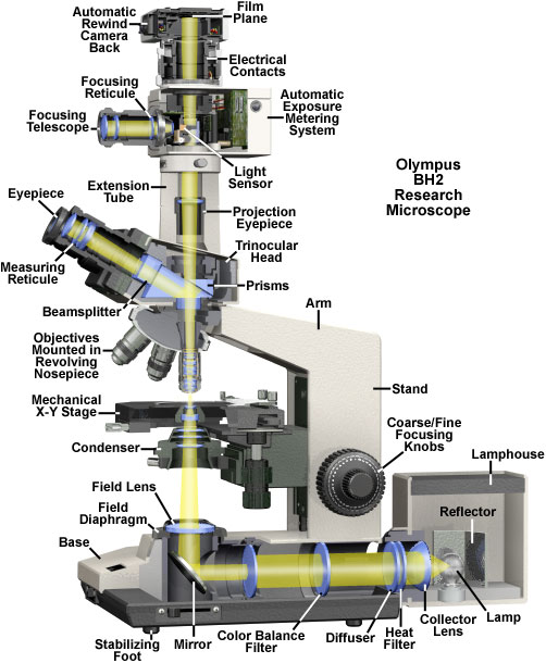
Olympus BH2 Research Microscope Cutaway Diagram | Olympus LS

Parts of a Microscope Labeling Activity

Parts of the Microscope with Labeling (also Free Printouts ...

File:Microscope diagram.png - Wikimedia Commons
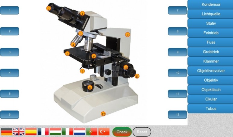
Aufbau Mikroskop lernen - Software kostenlos | Light Microscope

label microscope diagram | Charts | Microscope, Polarizing ...
Labelling Microscope - Labelled diagram

Compound Microscope- Definition, Labeled Diagram, Principle ...

Compound Microscope Parts – Labeled Diagram and their ...
0 Response to "38 labeled diagram of a microscope"
Post a Comment