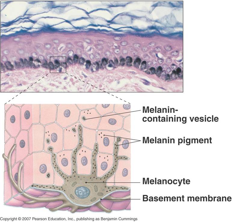
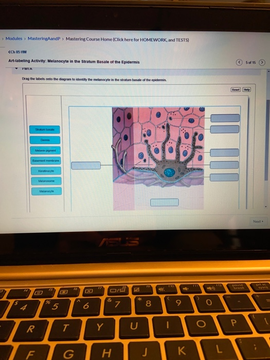
36 drag the labels onto the diagram to identify the melanocyte in the stratum basale of the epidermis.
Anatomy, Skin (Integument), Epidermis - StatPearls - NCBI ... Nov 19, 2021 · The layers of the epidermis include the stratum basale (the deepest portion of the epidermis), stratum spinosum, stratum granulosum, stratum lucidum, and stratum corneum (the most superficial portion of the epidermis). Stratum basale, also known as stratum germinativum, is the deepest layer, separated from the dermis by the basement membrane ... Cells of the Human Body - DocShare.tips Sebaceous lipids contribute to maintaining the integrity of the skin barrier, and express pro-inflammatory and anti-inflammatory properties.[10][][] Recent research suggests that sebum may represent a delivery system for antioxidants, antimicrobial lipids, pheromones, and hydration of stratum corneum.[]
Layers of the Skin - Anatomy and Physiology Stratum Basale. The stratum basale (also called the stratum germinativum) is the deepest epidermal layer and attaches the epidermis to the basal lamina, below which lie the layers of the dermis. The cells in the stratum basale bond to the dermis via intertwining collagen fibers, referred to as the basement membrane. A finger-like projection, or fold, known as the dermal papilla (plural ...

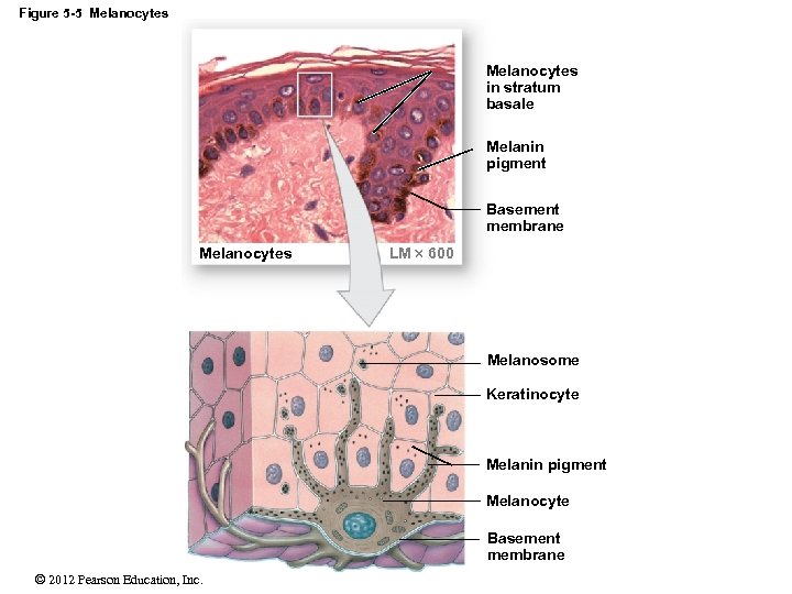
Drag the labels onto the diagram to identify the melanocyte in the stratum basale of the epidermis.
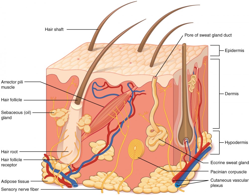
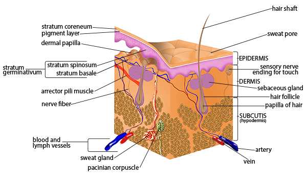
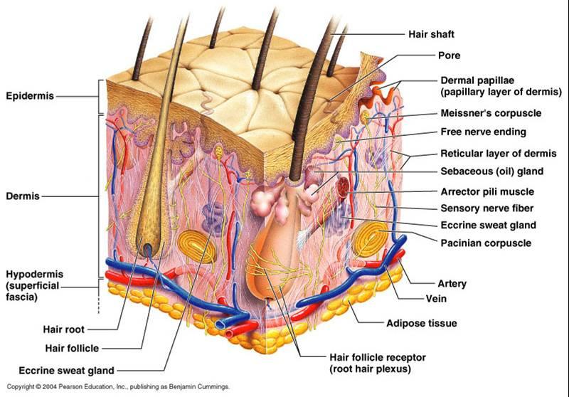
To learn the components of the integumentary system Label ... To learn the components of the integumentary system. Label the components of the integumentary system. Part A Drag the labels onto the diagram to identify the components of the integumentary system. ANSWER: Help Reset Stratum basale Dermis Basement membrane Melanocyte Melanin pigment Keratinocyte Melanosome. ANSWER : A&P Chapter 5 The Integumentary System ... - Easy Notecards melanin. 31. The most dangerous type of skin cancer is ________. melanoma. 32. The pinkish hue of individuals with fair skin is the result of the crimson color of oxygenated hemoglobin (contained in red blood cells) circulating in the dermal capillaries and reflecting through the epidermis. True. 33. Skin Anatomy: The Layers of Skin and Their Functions Feb 06, 2022 · The epidermis is made up of five individual layers: 2. Stratum basale: This bottom layer, also known as the basal cell layer, has column-shaped cells that push older cells toward the surface. As the cells move upward, they start to flatten and die. The layer is also made up of melanocytes (that produce a pigment that gives the skin its color ...
Drag the labels onto the diagram to identify the melanocyte in the stratum basale of the epidermis.. Ch 5-7 Lab A&P Mastering Flashcards - Quizlet Drag the labels onto the diagram to identify the cells and fibers of connective tissue proper using diagrammatic and histological views. ... Drag the labels onto the diagram to identify the basic structures of the epidermis-dermis junction. look at pic. Drag the labels onto the diagram to identify the melanocyte in the stratum basale of the ... Full text of "Cosmetic Dermatology Products And Procedures ... An icon used to represent a menu that can be toggled by interacting with this icon. Histology - Yale University Basal Cell Layer - Keratinocytes begin in the deepest layer of the epidermis, the stratum basale, which is a row of columnar cells resting on the basal lamina that separates the epidermis from the dermis. Mitosis occurs exclusively at the basal cell layer and allows for the replacement of cells lost from the surface. Human Anatomy & Physiology Laboratory Manual Main Version ... 4 Identifying Organs in the Abdominopelvic Cavity 8 ... • NEW! Drag-and-Drop Art Labeling Questions let students assess their knowledge of terms and structures. • Updated! Assignable pre-lab and post-lab quizzes for all 46 exercises in the lab manual. ... epidermis, dermis, and cutaneous sense organs 6. testis, ductus deferens, urethra 7.
Layers of the Skin | Anatomy and Physiology - Lumen Learning The stratum basale (also called the stratum germinativum) is the deepest epidermal layer and attaches the epidermis to the basal lamina, below which lie the layers of the dermis.The cells in the stratum basale bond to the dermis via intertwining collagen fibers, referred to as the basement membrane. The stratum basale is a single layer of cells primarily made of basal cells. 5.1 Layers of the Skin – Anatomy & Physiology Stratum Basale. The stratum basale (also called the stratum germinativum) is the deepest epidermal layer and attaches the epidermis to the basal lamina, below which lie the layers of the dermis. The cells in the stratum basale bond to the dermis via intertwining collagen fibers, referred to as the basement membrane. The 5 Layers of Your Skin - Dr. Leslie ... - Leslie Baumann 1. Stratum Basale or Basal Layer. The deepest layer of the epidermis is called the stratum basale, sometimes called the stratum germinativum. This is where stem cells are located. Because this layer is the innermost layer, many topical products that you apply to the surface of your skin cannot reach this layer and have an effect. Drag the labels onto the epidermal layers. Drag the labels ... Reset Help Stratum basale Stratum lucidum Dermis Dermal papilla Stratum corneum Basement membrane Stratum granulosum Epidermal ridge Stratum spinosum . ... Drag the labels onto the diagram to identify the layers of the epidermis. Reset Help stratum lucidun stratum comum stratum basale stratum spinosum 6. Name the specific sub-layer of the ...
Building a Medical Terminology Foundation The epidermis is composed of keratinized, stratified squamous epithelium. It is made of four or five layers of epithelial cells, depending on its location in the body. It is avascular. Thin skin has four layers of cells. From deep to superficial, these layers are the stratum basale, stratum spinosum, stratum granulosum, and stratum corneum ... Fundamentals of anatomy & physiology [Tenth edition ... The stratum basale and the underlying dermis interlock, strengthening the bond Epidermis between the two. The Basement epidermis forms epidermal membrane ridges, which extend into Epidermal ridge the dermis and are Dermal adjacent to dermal papilla papillae that project into Dermis the epidermis. Sympathetic Division (Thoracolumbar) Dermis Thin ... PDF Respondus LockDown Browser Installation Learning and Applied Innovation 8/19 . Respondus LockDown Browser. TM. Installation . Respondus LockDown Browser may be required to access exams in courses using Blackboard. a&p lab 7 hw Flashcards | Quizlet Drag the labels onto the diagram to identify the main structural features in the epidermis of thin skin. left column: dermis ... The cells in this layer of epidermis are dead, and their flat, scale-like remnants are filled with keratin. ... Skin cancer arising in the stratum basale is called basal cell CARCINOMA.
MAKE UP ARTIST HANDBOOK by pms1967 - Issuu
Integumentary System - Building a Medical Terminology ... Epidermis. The epidermis is composed of keratinized, stratified squamous epithelium. It is made of four or five layers of epithelial cells, depending on its location in the body. It is avascular.. Thin skin has four layers of cells. From deep to superficial, these layers are the stratum basale, stratum spinosum, stratum granulosum, and stratum corneum.Most of the skin can be classified as thin ...

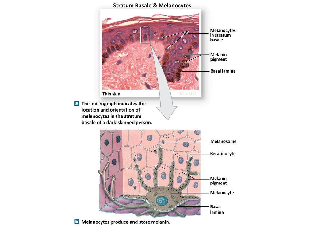
Art-labeling Activity: Melanocyte in the Stratum Basale of ...
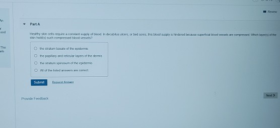
Part A Drag the labels onto the diagram to identify ... Anatomy and Physiology questions and answers. Part A Drag the labels onto the diagram to identify the melanocyte in the stratum basale of the epidermis. Reset Help Melanin pigment Basement membrane 20 Strahur basale III II Melanosome Demme Melanocyte Keratinocyte Reww an Part A od Healthy skin els require a constant supply of blood In decubus ...
5 The Integumentary System Power Point Lecture Presentations
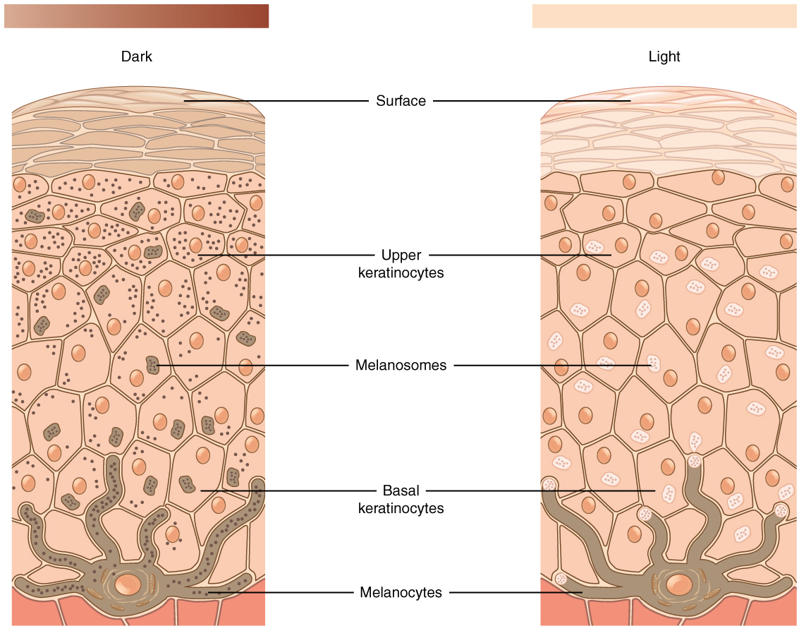
A. Tactile cells anchor the skin to the body. B. Keratinocytes produce a fibrous protein to protect the epidermis. C. Melanin provides protection against ultraviolet (UV) radiation. D. Langerhans cells activate the immune system. A. Tactile cells anchor the skin to the body. The skin consists of two main regions.
Layers of the Skin | Anatomy and Physiology I
Solved Drag the labels onto the diagram to identify the ... Transcribed image text: Modules MasteringAandP Mastering Course Home (Click here for HOMEWORK, and TESTS) Ch 05 HW Art-labeling Activity: Melanocyte in the Stratum Basale of the Epidermis 5 of 15 rart A Drag the labels onto the diagram to identity the melanocyle in the stratum basale of the epidermis Reset Helo Statum basae Demis Melann pgment Basement mentrane Keratinocye Melanosome Melaocyle ...
Layers of the Skin – Anatomy and Physiology
(PDF) Anatomy | Mohamed Mahdi - Academia.edu Enter the email address you signed up with and we'll email you a reset link.
AP Biology
Keratinocytes in the stratum lucidum start producing ... 10/14/2016 API Lab Homework 6 6/9 Drag the labels onto the diagram to identify the melanocyte in the stratum basale of the epidermis. ANSWER: Correct Chapter 5 Clinical Note Questions 1 and 2: Skin Cancer Skin Cancer Almost everyone has several benign tumors of the skin. Moles and warts are common examples. However, skin cancers, which are more dangerous, are the most common form of cancer.
SEER Training: Anatomy of the Skin
Modern Technology Of Cosmetics by Niir Board, ISBN ... Stratum granulosum The next layer within the epidermis is the stratum granulosum or granular layer. As cells progress to the surface, they form the characteristic granules of this layer. ... Melanin is produced by the melanocyte cells present in the stratum basale. These melanocytes are present in this basal layer in great abundance and the ...
Associate Degree Nursing Physiology Review
PDF Label The Skin Anatomy Diagram Answers The cells in all of the layers except the stratum basale are called keratinocytes. A keratinocyte is a cell that manufactures and ... melanocyte - a cell in the epidermis that produces circulates. ... label with a line or put the label directly onto the area described. Be as precise as possible. If you are worried about the precision
The Art and Science of Dermal Formulation Development
pubmed.ncbi.nlm.nih.gov pubmed.ncbi.nlm.nih.gov
Untitled
Epidermis - 5 Layers of Epidermis, Outermost Layer & Function The epidermis of thick skin has five layers. Beginning at the basal lamina and traveling superficially toward the epithelial surface, we find the stratum basale, stratum spinosum, stratum granulosum, stratum lucidum, and stratum corneum. Refer to Figure 2 as we describe the layers in a section of thick skin.

Nanoparticles influence in skin penetration of drugs: In ...
(PDF) Anatomy, Histology & Cell Biology ... - Academia.edu Anatomy, Histology & Cell Biology PreTest Self-Assessment & Review (4th Ed.)[vickey]
A&P Chapter 5 The Integumentary System Flashcards - Easy ...
Art-labeling Activity: Melanocyte in the Stratum Basale of ... Start studying Art-labeling Activity: Melanocyte in the Stratum Basale of the Epidermis. Learn vocabulary, terms, and more with flashcards, games, and other study tools.

2-1 Mastering AP Lab - Module Two Homework.docx - 2-1 ...
Skin Anatomy: The Layers of Skin and Their Functions Feb 06, 2022 · The epidermis is made up of five individual layers: 2. Stratum basale: This bottom layer, also known as the basal cell layer, has column-shaped cells that push older cells toward the surface. As the cells move upward, they start to flatten and die. The layer is also made up of melanocytes (that produce a pigment that gives the skin its color ...

LAB A&P Flashcards | Quizlet
A&P Chapter 5 The Integumentary System ... - Easy Notecards melanin. 31. The most dangerous type of skin cancer is ________. melanoma. 32. The pinkish hue of individuals with fair skin is the result of the crimson color of oxygenated hemoglobin (contained in red blood cells) circulating in the dermal capillaries and reflecting through the epidermis. True. 33.
The Integumentary system - ppt download
To learn the components of the integumentary system Label ... To learn the components of the integumentary system. Label the components of the integumentary system. Part A Drag the labels onto the diagram to identify the components of the integumentary system. ANSWER: Help Reset Stratum basale Dermis Basement membrane Melanocyte Melanin pigment Keratinocyte Melanosome. ANSWER :

Drag the labels onto the epidermal layers. Drag the labels ...

Ch4 skin, martini
Vieillissement cutané Skin ageing

Anatomy This document is attributed to: J. Gordon Betts, Tyler ...
The Integumentary System

5 The Integumentary System. - ppt download
Sample report - Filter (14041 patents) - PatentInspiration

PDF) Cosmetic Formulation Principles and Practice | Nadia ...

LAB A&P Flashcards | Quizlet
Exercise 7 Flashcards - Easy Notecards

Welcome to the Open Learning Initiative Introduction to ...
Muscular System, Chapter 10
Concise Histology

5.1 Layers of the Skin – Anatomy & Physiology
The Integumentary System
Genital sistem

What Is Melanoma Skin Cancer? | What Is Melanoma?
Unit 4
Part A Drag the labels onto the diagram to identify | Chegg.com
Solved Drag the labels onto the diagram to identify the ...

The layer of epidermis that is the closest to the dermis is ...
0 Response to "36 drag the labels onto the diagram to identify the melanocyte in the stratum basale of the epidermis."
Post a Comment