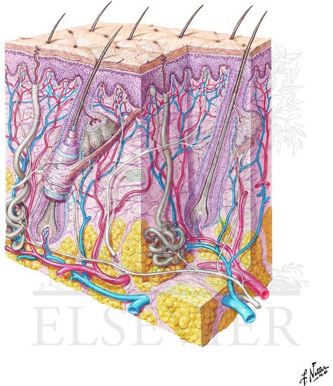
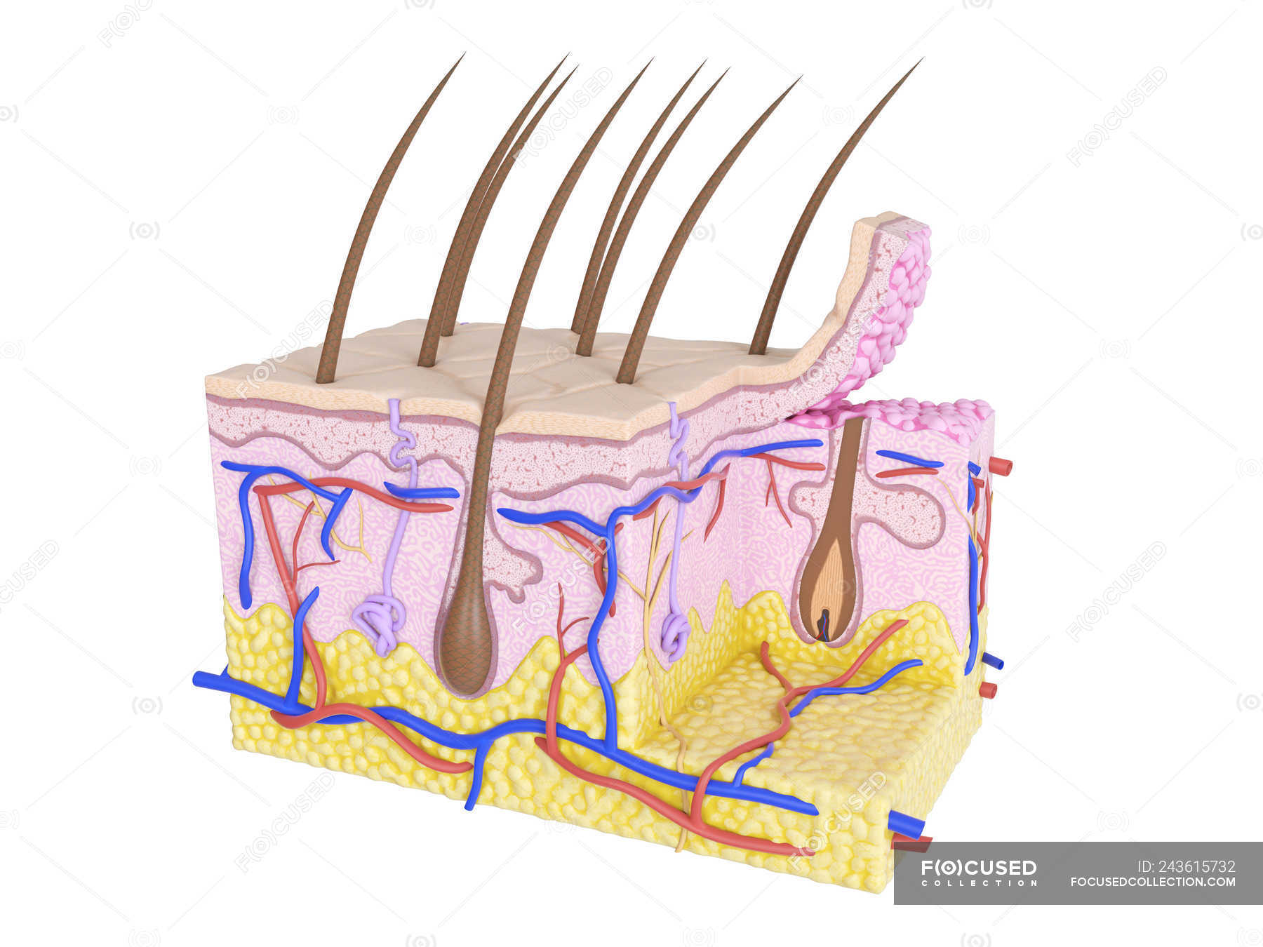
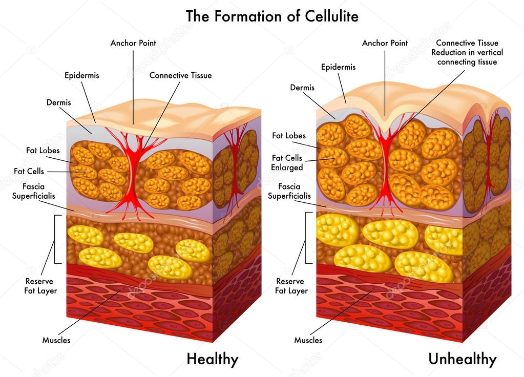
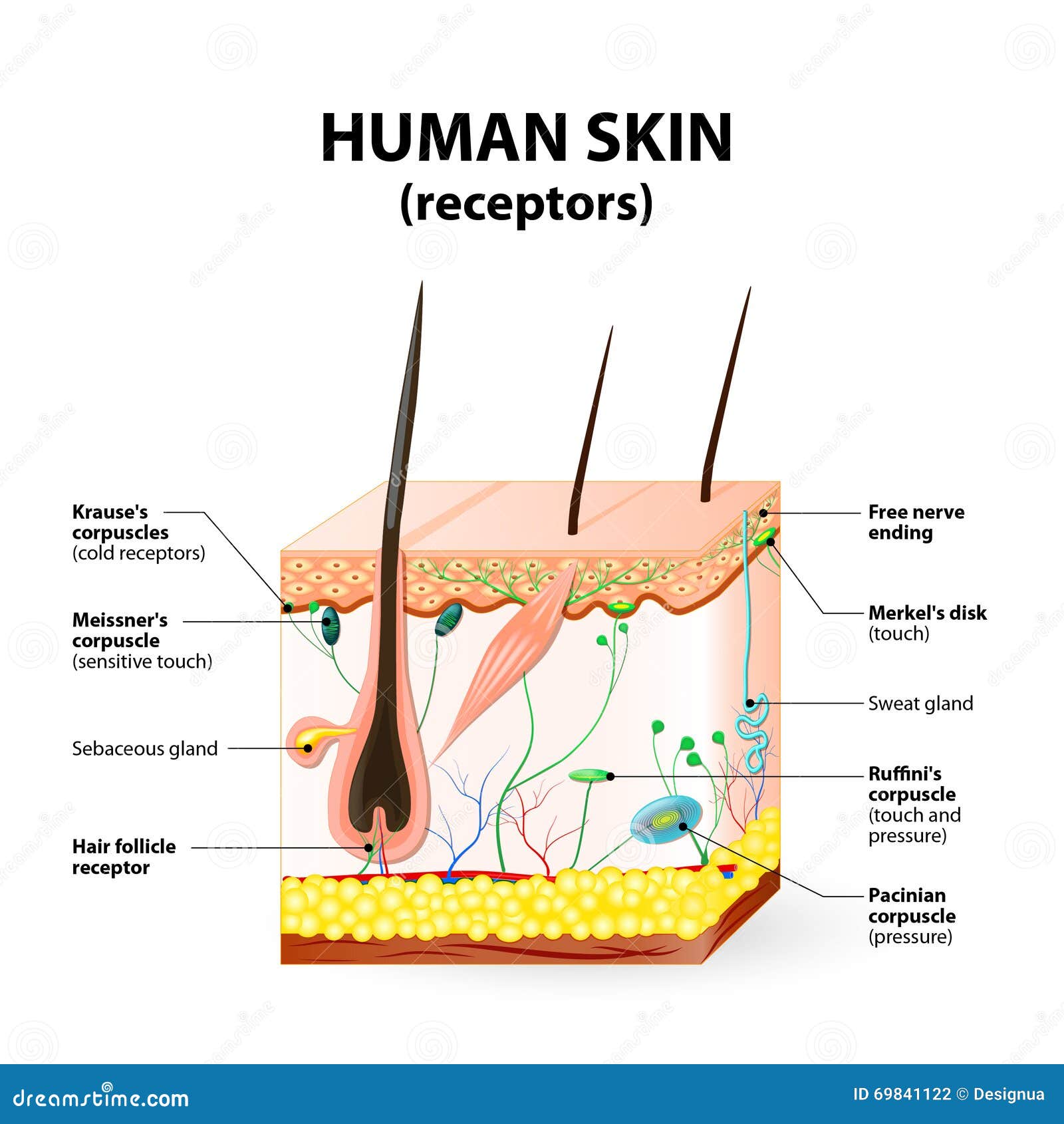
40 skin cross section diagram
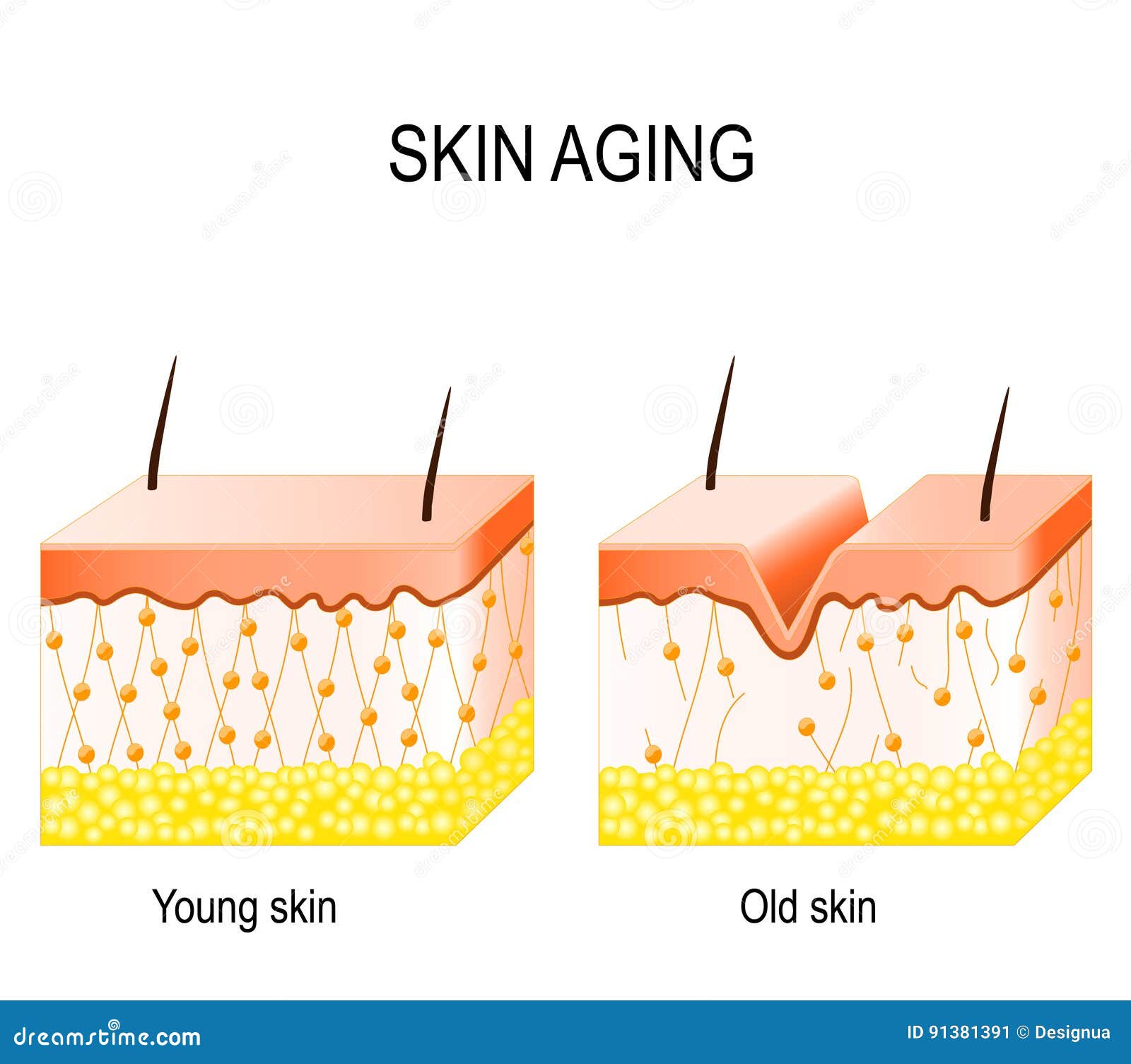
Summary. Skin is the protective covering of our body that appears to be a thin sheet. A cross-section through the skin explains that the skin of humans and other mammals is primarily composed of three distinct layers. The outermost layer is the epidermis followed by the dermis and hypodermis. Illustration about Aging skin. Cross section young and old skin. the diagram showing the decrease in collagen and broken elastin in older skin. Illustration of science, elastin, diagram - 91381391
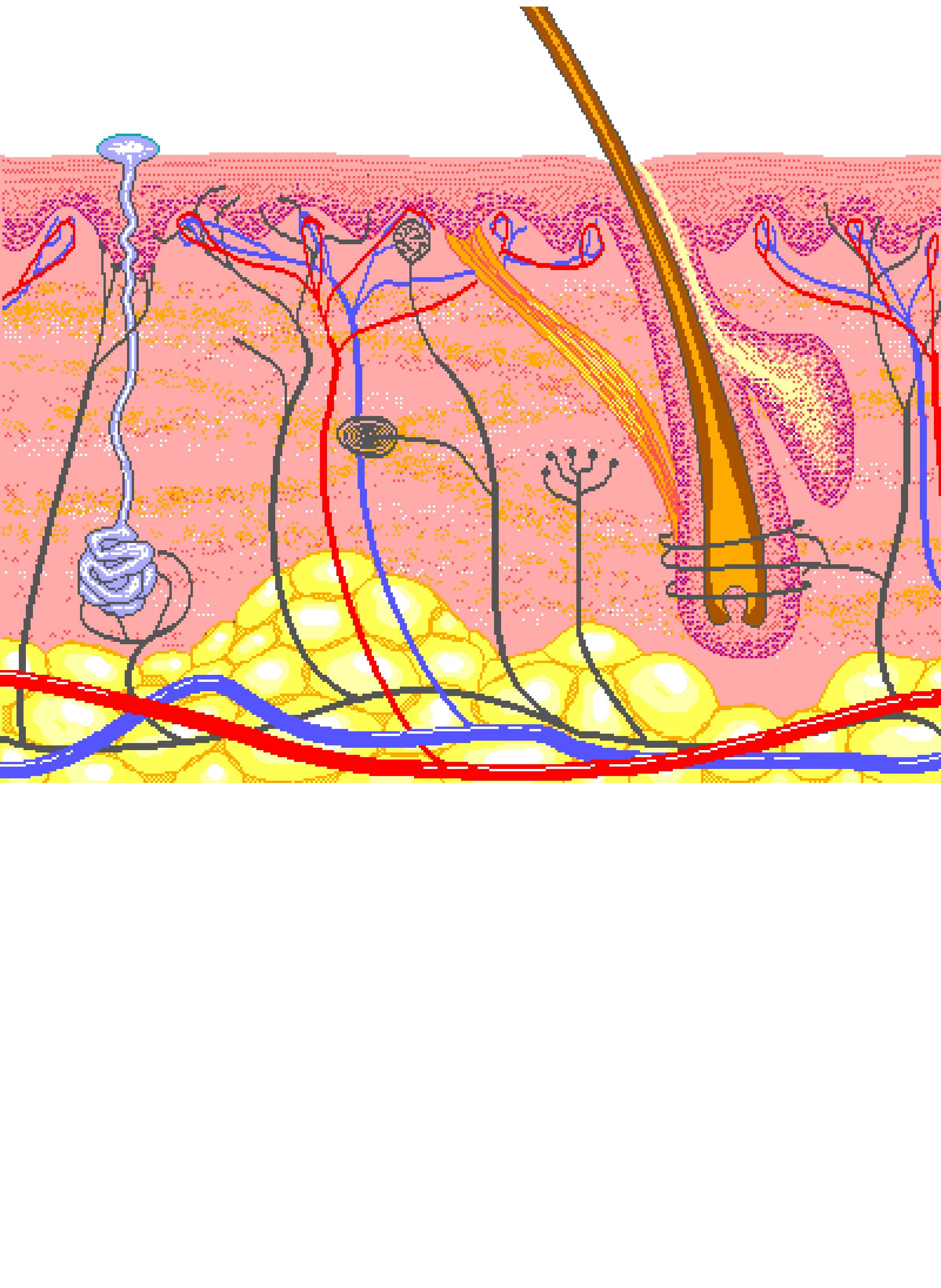
Look at the cross section diagram of the skin below. Choose the best answers to fill in each of the missing labels. epidermis. nerve. fat. hair. sebaceous gland. capillary. dermis. muscle . 2. In the following sentences, choose the best answer to fill each of the blanks. microorganisms. hair. protects. capillaries. epidermis.

Skin cross section diagram
Start studying Skin Cross-Section Integumentary System. Learn vocabulary, terms, and more with flashcards, games, and other study tools. by S Lawton · 2019 · Cited by 19 — The layer below the dermis, the hypodermis, consists largely of fat. These structures are described below. Fig-1-Cross-section-through-the-skin- ... This illustration shows a cross section of skin tissue. ... These slides show cross-sections of the epidermis and dermis of (a) thin and (b) thick skin.
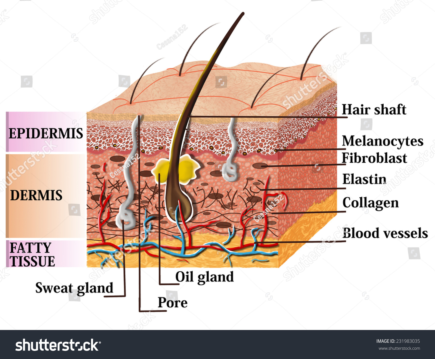
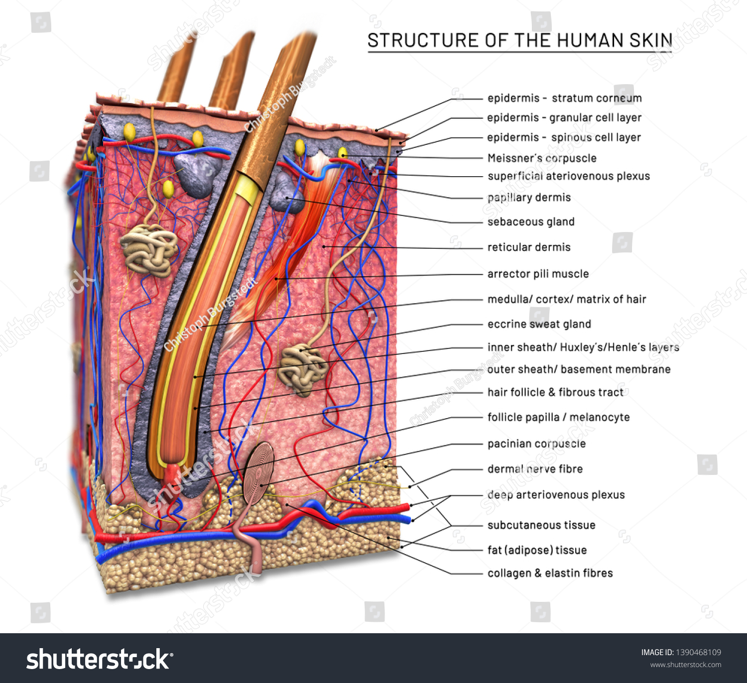
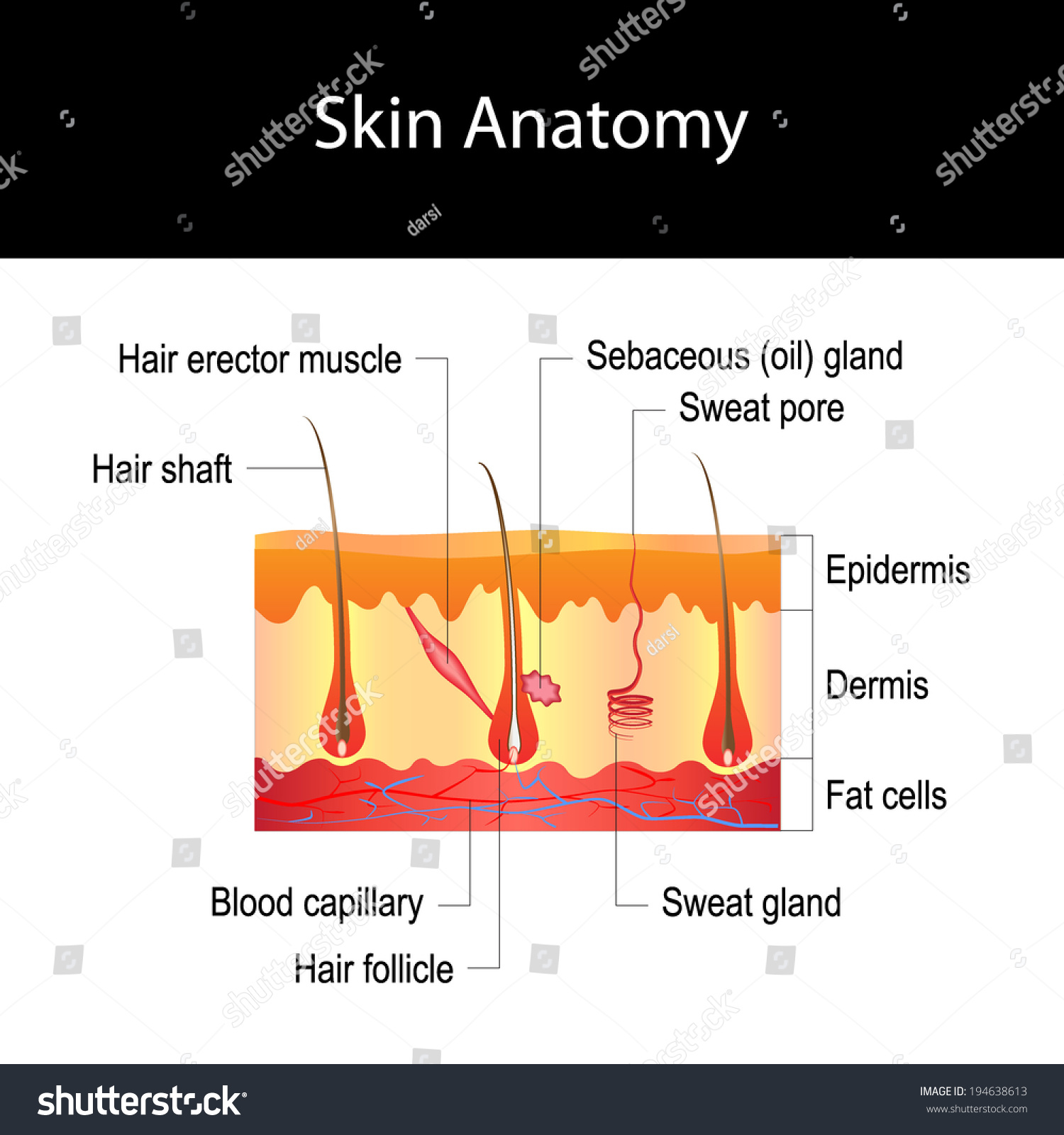
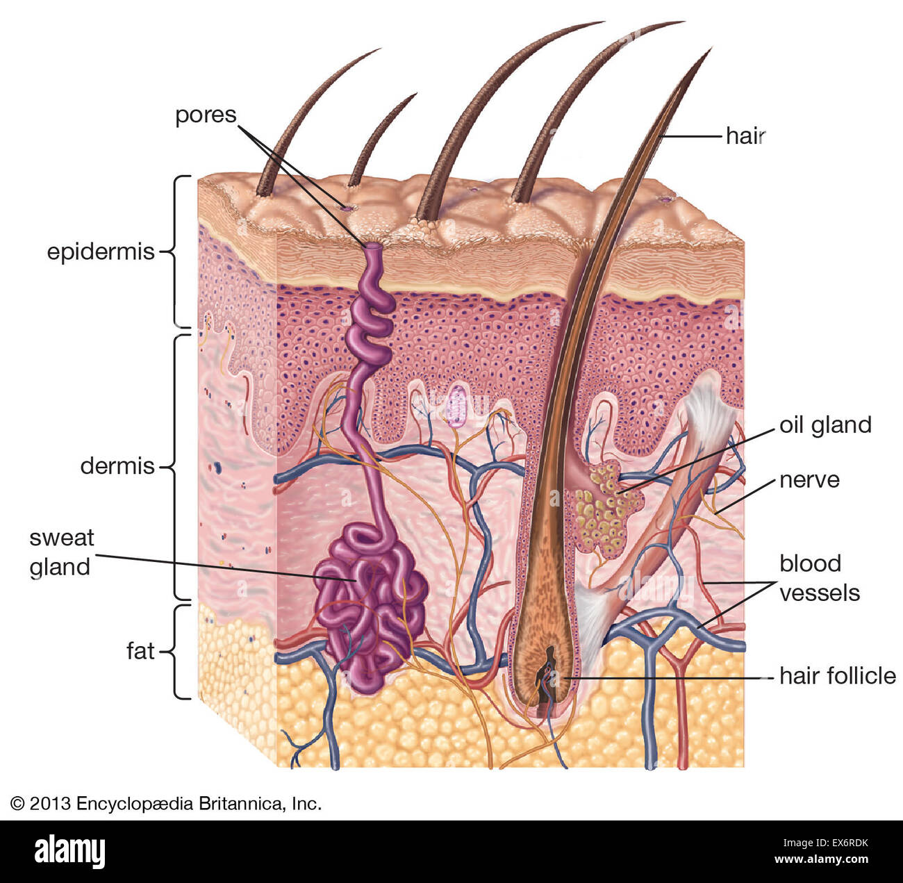
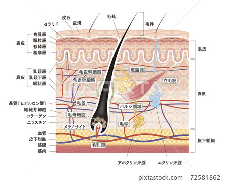
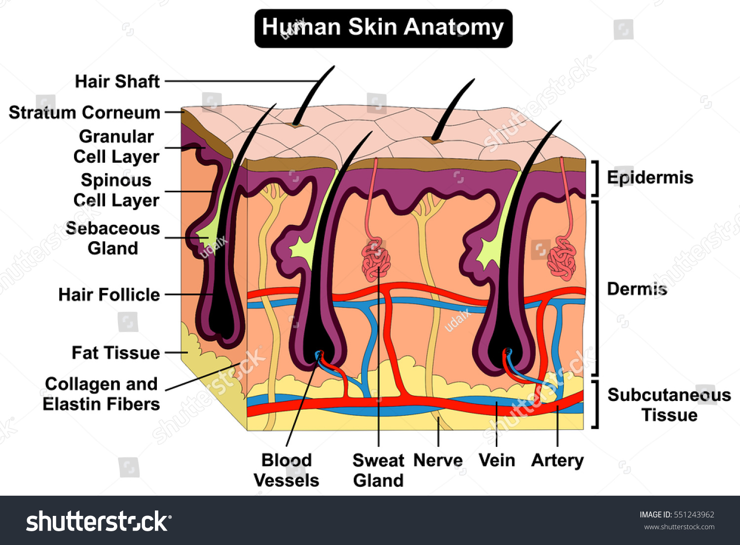
Skin cross section diagram. Browse 522 epidermis diagram stock photos and images available, or search for cross section skin or skin layers to find more great stock photos and pictures. Psoriasis in section, skin with detailed epidermis in stratum corneum, granular and germinative, dermis, and hypodermis. Skin is a waterproof, flexible, but tough protective covering for your body. Normally the surface is smooth, punctuated only with hair and pores for sweat. A cross-section of skin shows the major parts. It is divided into three layers. The outer layer is the epidermis. The dermis is in the middle and fat forms the innermost layer. Find Human Skin Anatomy Cross Section Diagram stock images in HD and millions of other royalty-free stock photos, illustrations and vectors in the Shutterstock collection. Thousands of new, high-quality pictures added every day. "Thick skin" is found only on the palms of the hands and the soles of the feet. It has a fifth layer, called the stratum lucidum, located between the stratum corneum and the stratum granulosum (Figure 5.1.2). Figure 5.1.2 - Thin Skin versus Thick Skin: These slides show cross-sections of the epidermis and dermis of (a) thin and (b) thick ...
Human Skin Cross Section Anatomy Diagram. Human skin cross section anatomy including all parts for medical science education and health care ... Human Tooth Cross Section Anatomy Diagram including enamel dentine pulp cavity gum tissue bone nerve blood vessels cement canal part crown neck root teeth types incisors canine molars dental for ... Download scientific diagram | A schematic cross-section of human skin [9]. from publication: Prodrug Strategies for Enhancing the Percutaneous Absorption of ... The best selection of Royalty Free Skin Cross Section Diagram Vector Art, Graphics and Stock Illustrations. Download 100+ Royalty Free Skin Cross Section Diagram Vector Images. Download scientific diagram | A diagrammatic representation of the structure of human skin in cross section. The epidermis is composed of the stratum corneum and the viable epidermis. Diagram is ...
There are two main skin layers: The outer layer (epidermis); The inner layer (dermis). The skin cells (melanocytes) that develop into melanoma usually are ... Oct 23, 2015 - Find Cross Section Human Skin Labels stock images in HD and millions of other royalty-free stock photos, illustrations and vectors in the Shutterstock collection. Thousands of new, high-quality pictures added every day. 8,623 skin cross section stock photos, vectors, and illustrations are available royalty-free. See skin cross section stock video clips. of 87. scalp anatomy pigment hair skin vessels cross section of veins hair follice muscle labelled structure of the hair follicle layers epidermis hair anatomy hair section. Try these curated collections. Find the perfect human skin diagram stock photo. ... Skin anatomy diagram concept with a cross section of the human body surface organ with hair follicle ...
Jul 3, 2020 - Illustration about Cross section of skin showing all layers and major appendages. Illustration of shaft, cross, skin - 9845525
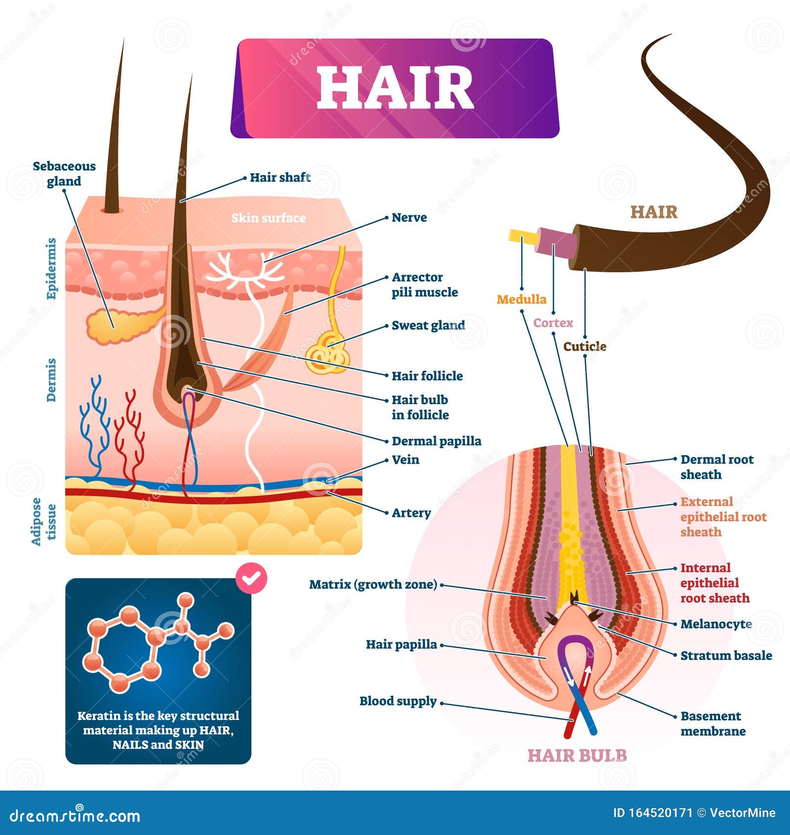
Diagram of a hair follicle in a cross section of skin layers. Illustration about cortex, medulla, away, human, care, capillary, health, healthy, growth, layer ...
Magnification: x14,000. TEM of several red cells in cross-section within the capillary loop (any of the small blood vessels that carry blood in the papillae of the skin) of a glomerulus from the kidney of a mouse. The hemoglobin-containing red cells appear deeply gray and homogeneous because they contain no nuclei.
Diagram showing a cross section of skin on the left and on the right a cross section showing the cell types. Appears in. ARTICLE. Skin structure. The epidermis is the thin layer at the surface that varies in thickness from 0.05 mm on the eyelids to 1.5 mm on the palms of the feet. The top of the epidermis is called the cornified layer, and ...
Download scientific diagram | Schematic drawing of cross-section of the human skin. from publication: Visualization of Epidermis and Dermal Cells in ex vivo Human Skin Using the Confocal and Two ...
Cross section through the thalamus: Diagram Orienting yourself within such a cross section is easy. The star of the show (brain) is easily recognizable because it appears highly convoluted, full of ridges (gyri) and indentations (sulci).The paired thalami appear as two circular masses in the midline, forming the walls of the third ventricle.The neurocranium appears as a meshwork (trabecular ...
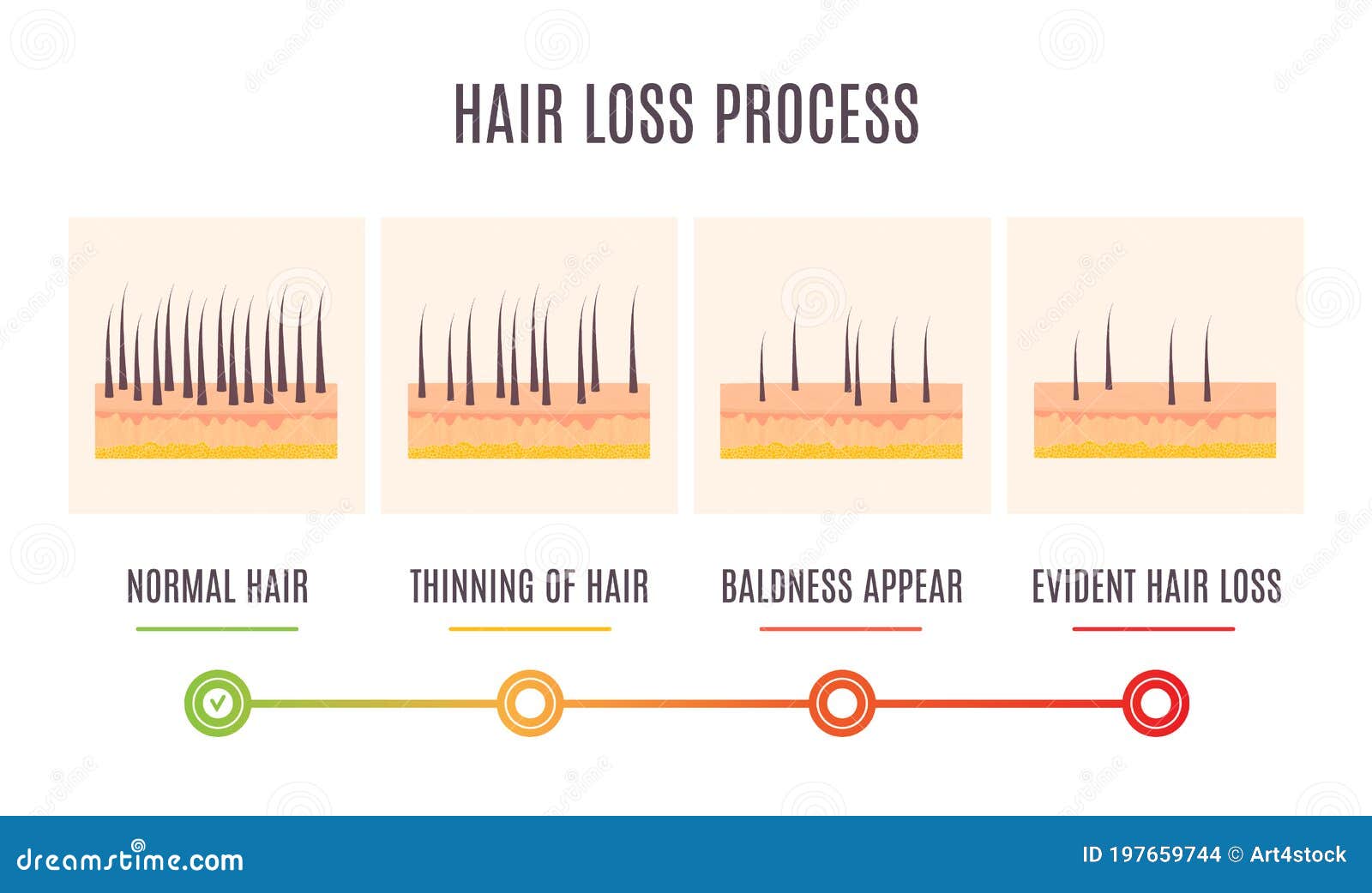
Male pattern baldness set with skin cross-section diagram. Treatment result in top view. Alopecia infographics medical vector template for clinics.
Skin Cross-Section. The skin is by far the largest organ of the human body, weighing about 10 pounds (4.5 kg) and measuring about 20 square feet (2 square meters) in surface area. It forms the outer covering for the entire body and protects the internal tissues from the external environment. The skin consists of two distinct layers: the ...
Browse 549 skin diagram stock photos and images available, or search for aging skin diagram to find more great stock photos and pictures. Psoriasis in section, skin with detailed epidermis in stratum corneum, granular and germinative, dermis, and hypodermis.
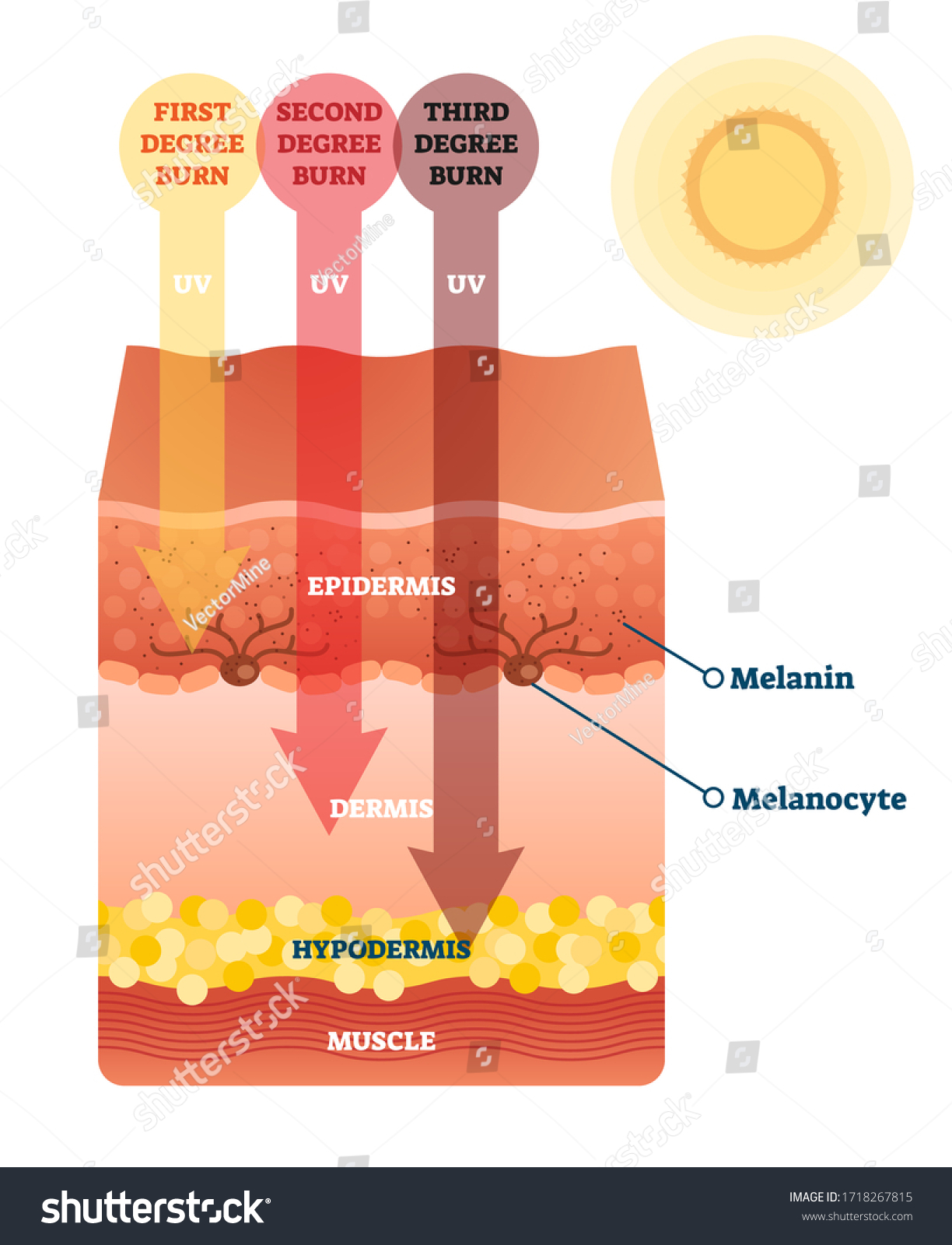
Skin also helps maintain a constant body temperature. Human skin is only about 0.07 inches (2 mm) thick. Skin is made up of two layers that cover a third fatty layer. The outer layer is called the epidermis; it is a tough protective layer that contains melanin (which protects against the rays of the sun and gives the skin its color).
Skin Poster - Medical Anatomy Poster - 24" x 36" Laminated Quick Reference. This skin covers a lot of area. This Skin poster chart vividly describes and illustrates layers of the epidermis, skin area, nail structures, pimples, burn classification, hair follicles, and the Rule of Nines. liztran5188. L.
Vector image "Diagram of a hair follicle in a cross section of skin layers" can be used for personal and commercial purposes according to the conditions of the purchased Royalty-free license. The illustration is available for download in high resolution quality up to 5806x4376 and in EPS file format.
The Skin - Science Quiz: The skin is an organ. In fact, it's the body's largest organ and is responsible for protection against germs that can cause infection. It also gives us our sense of touch and helps control the body's temperature. The skin is an amazing part of the human body, and this science quiz game will help you identify its components. The middle section of the skin is ...
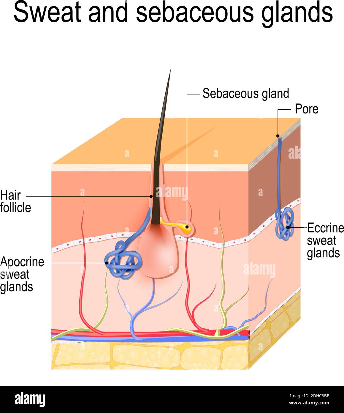
Skin Anatomy Skin anatomy diagram concept with a cross section of the human body surface organ with hair follicle and red and blue blood vessels as a health care and medical symbol of anatomical function. skin cross section stock pictures, royalty-free photos & images
Part 1: Skin 1. View the diagram of skin in cross section below. Use it as a reference in this Part, as needed. Skin Cross Section Diagram 2. Take the skin cross section slide from the Containers shelf and place it on the microscope stage. 3. Observe the epidermis. You may need to use the y-axis stage adjust knob to locate it.
Structure of the humans skin. Vector diagram for your design, educational, science and medical use skin cross section diagram drawing stock illustrations. Vector illustration of human hair diagram. Piece of human skin and all structure of hair on the white background. Medical Treatment of baldness, epilation concept.
Start studying Chapter 1: Skin Cross Section. Learn vocabulary, terms, and more with flashcards, games, and other study tools.
This illustration shows a cross section of skin tissue. ... These slides show cross-sections of the epidermis and dermis of (a) thin and (b) thick skin.
by S Lawton · 2019 · Cited by 19 — The layer below the dermis, the hypodermis, consists largely of fat. These structures are described below. Fig-1-Cross-section-through-the-skin- ...
Start studying Skin Cross-Section Integumentary System. Learn vocabulary, terms, and more with flashcards, games, and other study tools.















0 Response to "40 skin cross section diagram"
Post a Comment