36 cat muscles labeled diagram
muscle,you will have to cut the superficial muscle at the midline and reflect (pull back) the edges toward the origin and insertion. B. Muscles of the Head and Neck 1. Refer to Figure C1.1 to locate the following superfi-cial muscles on the cat. Cats have a platysma,but this muscle was most probably removed during the skin-ning process ... The muscles of the cat are similar to the muscles on a human. The pictures below identify major muscle groups in the cat. Some are labeled and some are not labeled (for use in slide tests). Anatomy Corner resource site for teachers and students of Anatomy and Physiology. Find quizzes, diagrams, and slide presentations on structures, functions ...
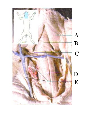
The following links will allow you to access real photographs of the cat muscular system. The purpose of these pages is to quiz your knowledge on the structures of the muscular system. Please try to answer all structures (or guess) before you look at the answers! Choose one of the following categories: Neck Muscles. Neck (Superficial)

Cat muscles labeled diagram
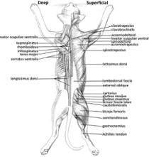
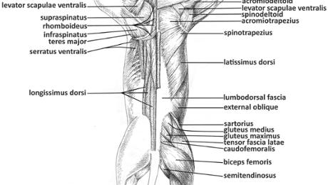
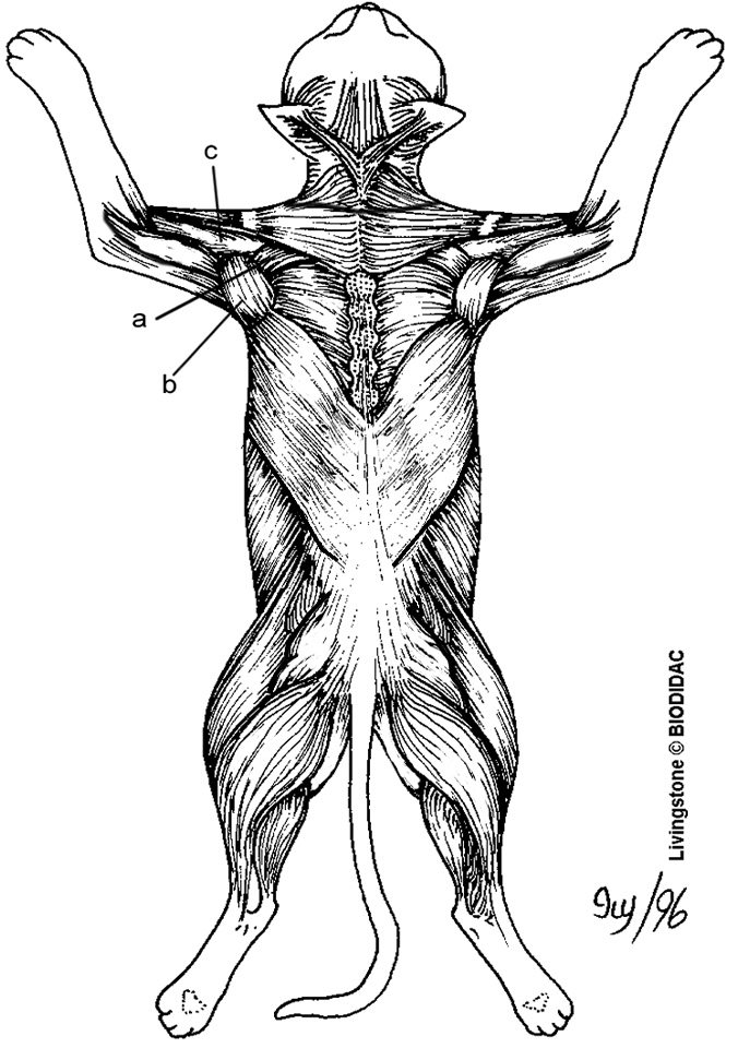
Muscle Lab 1 The study of cat musculature starts on p.309 of the lab manual. First sex your cat. Read over the section "Preparing the cat for muscle dissection" on p. 310. Since the pictures in your manual tend to be only close-ups, I have provided some diagrams that show the location of all the Cat Anatomy Dissection Guide Superficial Muscles Ventral View pectoantebrachialis Dorsal View clavotrapezius pectoralis major acromiotrapezius pectoralis minor spinotrapezius xiphihumeralis latissiumus dorsi external oblique clavobrachialis internal oblique acromiodeltoid transversus abdominis spinodeltoid ... VCD - Muscles. The muscles of the cat are organized into major groups. For the purpose of a basic anatomy class, only the easily visible superficial muscles will be looked at. Muscles are held together by connective tissue called fascia, which appears white in color on the specimens. The fascia also serves as a boundary marker between muscles.
Cat muscles labeled diagram. Cross-sectional labeled anatomy of the head and neck of the domestic cat on CT imaging (bones of the skull, cervical spine, mandible, hyoid bone, muscles of the neck, nasal cavity and paranasal sinuses, oral cavity, larynx) The cat's third eyelid is known as the nictitating membrane. It is located in the inner corner of the eye, which is also covered by conjunctiva. In healthy cats, the conjunctiva of the eyelids is not readily visible and has a pale, pink color. Integumental. The two main integumentary muscles of a cat are the platysma and the cutaneous maximus. Anatomy. Muscles. cat. BIOL-2401. Hind Leg. Games by same creator. Sarcomere Quiz 24p Image Quiz. Scapula Quiz 12p Image Quiz. Histology - Cross section of vertebra with spinal cord 31p Image Quiz. Hand Quiz 16p Image Quiz. Femur Quiz 25p Image Quiz. ... This online quiz is called Cat Hind Legs Muscle Quiz. Cat Muscles (Images) 53 terms. meliciementor. Identify the Gender - 3rd declensions. 87 terms. Yonatan_Gut. RAD105-1 Facial Pictures. 75 terms. SithDoc89.
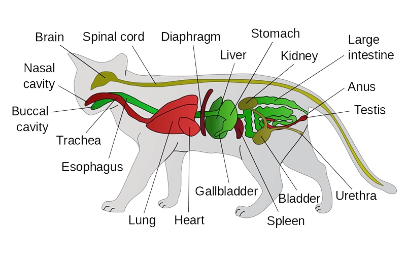
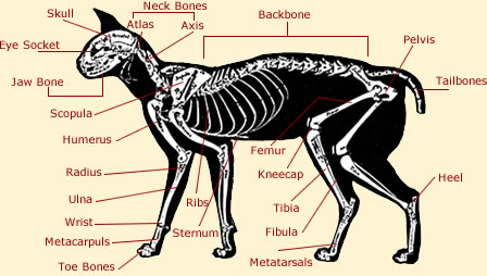
Basic feline anatomy. The following two diagrams help you familiarize yourself with basic feline anatomy. The chart below (of a male cat) shows you were all the internal organs are located. Did you know that cats have 244 bones in their body? Humans only have 206. This diagram of a feline skeleton shows you where all of your cat's bones are ... To play this quiz, please finish editing it. INSTRUCTOR-LED SESSION. Start a live quiz. SUPER. Classic. Students progress at their own pace and you see a leaderboard and live results. Instructor-paced BETA. Control the pace so everyone advances through each question together. Cat muscles are one of the essential parts of feline anatomy. Muscles—skeletal, cardiac and smooth—are tissues that contract to allow for movement and force. Muscles can make a cat travel from one place to another or make organs function properly. Skeletal Muscles, Cardiac Muscles and Smooth Muscles ... Unit 21: Anatomy of the Domestic Cat 1. Muscles of the cat abdomen 1 2. Muscles of the cat abdomen 2 3. Muscles of the cat abdomen 3 4. Superficial muscles of the cat chest 5. Muscles of the back and shoulder of the cat 6. Muscles of the lateral thigh of the cat 7. Muscles of the medial thigh of the cat 1 ...
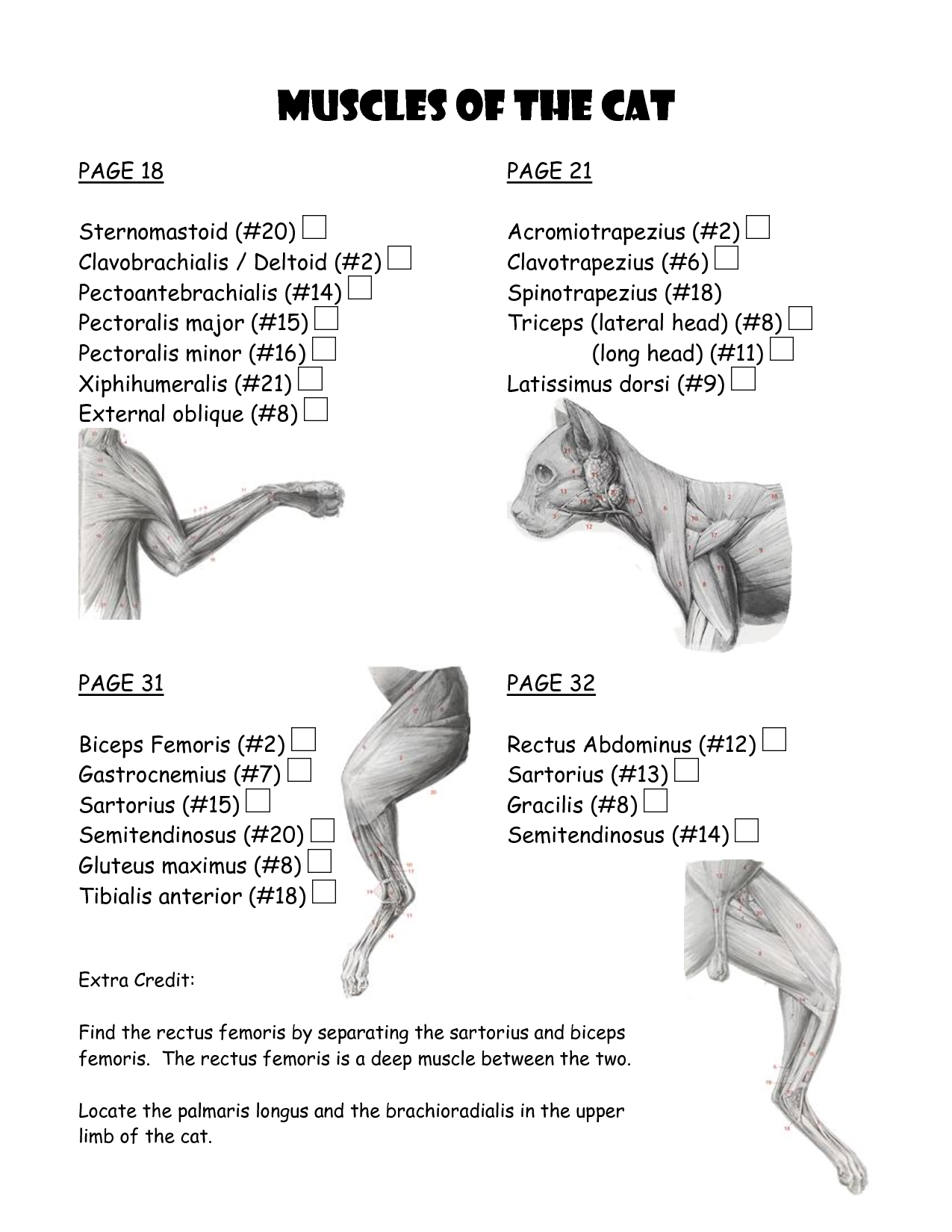
A human has 206 bones, however a cat has around 290 bones and 517 separate muscles, this makes them very agile animals, they use more than 500 muscles to leap, jump and sprint. A cat can jump over 7 times its own height. A cat has 13 ribs in its body. Take a look below at the diagram of a cats skeleton. The message makes the muscles contract (get smaller) or relax, which moves the bones of the skeleton and so the cat moves. Most of the cat's muscles are able to contract very quickly. This means the cat moves fast and can jump long distances. The cougar is known as a great jumper. Two of this cat's muscles are shown in the picture. Muscles of the cat : simplebooklet.com 1. Cat Dissection Muscular Labs. 2. External oblique Pectroalis minor Pectoralis major Gastrocnemius Sartorius Tibialis anterior Gracilis. 3. Latissimus dorsi Levator scapula Lumbodorsal fascia Gluteal muscles Deltoid Biceps femoris Gastrocnemius Trapezius Sartorius Trapezius Semitendinosis External oblique Deltoid Tensor fasciae latae Trapezius.
Browse 500 sets of quiz cat muscles flashcards. Study sets Diagrams Classes Users. 31 Terms. tallmadgiraffe. Cat Muscles. pectoralis major. pectoralis minor. rectus abdominis. external oblique.
This is an online quiz called Cat Muscle Anatomy. From the quiz author. name the muscels in a cats back Your Skills & Rank. Total Points. 0. Get started! Today's Rank--0. Today 's Points. One of us! Game Points. 14. You need to get 100% to score the 14 points available. Actions. Add to favorites 7 favs. Add to Playlist.
The cat skeletal anatomy labeled diagrams that provide in this article might help you a lot. But if you need more cat anatomy labeled diagrams, please let me known. Categories Dog and Cat Anatomy Tags cat skeleton , cat skeleton anatomy , cat skeleton anatomy labeled diagram , cat skeleton diagram , cat skeleton head , cat skeleton labeled ...
Anatomy & Physiology Cat Muscles ... The deepest lateral muscles of the cat shoulder, brachium, and forearm. Supraspinatus Infraspinatus Teres major deltoideus Triceps brachii long head Triceps brachii lateral head Brachialis . Sartorius Sartoñus (cut)
Find the rectus femoris by separating the sartorius and biceps femoris. The rectus femoris is a deep muscle between the two. Locate the palmaris longus and the brachioradialis in the upper limb of the cat.
Muscles are how we move and live. All movement in the body is controlled by muscles. Some muscles especially 5 Cat Muscle Anatomy Diagram work without us thinking, like our heart beating, while other muscles are controlled by our thoughts and allow us to do stuff and move around. There are over 650 muscles in the human body.
The digestive system ( cat) ( dog) includes the mouth, teeth, salivary glands, esophagus, stomach, intestine, pancreas, liver and gall bladder. The digestive system absorbs and digests food and eliminates solid wastes from the body. The integumentary system is the skin and fur that cover the animal's body. The skin protects the underlying organs.
Cat References Day 1 Terminology & External/Internal Anatomy. Pictures from iBook; Cat Skeleton Tutorial-Kenyon College; Day 2&3 Muscular System. Muscle Tables; Cutaneous, Shoulder & Back Muscles
Cat neck muscles anatomy
Cat Muscles of the Back (color) Image shows the dorsal (back) side of a cat with well defined muscles. Color the muscles to help learn them. Muscles include the trapezius, latissimus dorsi, gluteus maximus, triceps, biceps, gastrocnemius, and sartorius. tasco428.
Gain a comprehensive understanding of a cat's health with our veterinary guide to cat anatomy complete with diagrams, videos and simple explanations - cardiovascular, digestive, musckuloskeletal, respiratory, urogenital systems.
VCD - Muscles. The muscles of the cat are organized into major groups. For the purpose of a basic anatomy class, only the easily visible superficial muscles will be looked at. Muscles are held together by connective tissue called fascia, which appears white in color on the specimens. The fascia also serves as a boundary marker between muscles.
Cat Anatomy Dissection Guide Superficial Muscles Ventral View pectoantebrachialis Dorsal View clavotrapezius pectoralis major acromiotrapezius pectoralis minor spinotrapezius xiphihumeralis latissiumus dorsi external oblique clavobrachialis internal oblique acromiodeltoid transversus abdominis spinodeltoid ...
Muscle Lab 1 The study of cat musculature starts on p.309 of the lab manual. First sex your cat. Read over the section "Preparing the cat for muscle dissection" on p. 310. Since the pictures in your manual tend to be only close-ups, I have provided some diagrams that show the location of all the









0 Response to "36 cat muscles labeled diagram"
Post a Comment