39 correctly label the items in this dna replication fork diagram.
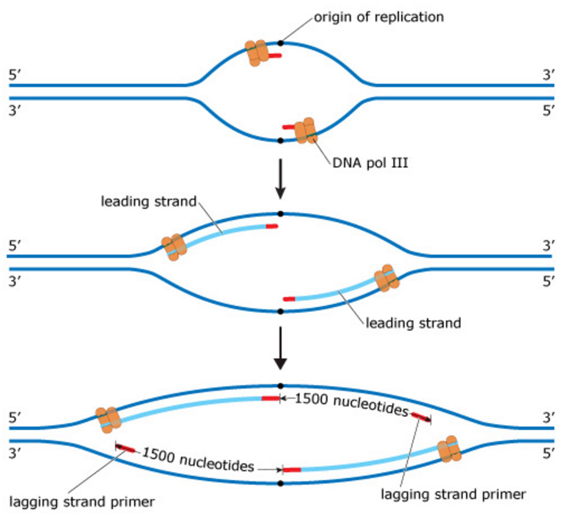
must be made in fragments (okazaki fragments) as the replication fork unwinds dna polymerase adds new nucleotides away from the fork. the fragments then need to be covalently bonded to make it a continuous complete strand H. Describe what a replication bubble is and label the replication bubbles in the diagram below Label DNA and Replication . ... SSBs Label the diagram: 5. DNA polymerase adds nucleotides (5' to 3') Replication fork is formed DNA polymerase attaches to the primer Okazaki fragments bound by ligase DNA helicase unwinds DNA Rearrange the steps to indicate the correct order: 6. 1. Enzyme that unwinds DNA 2.
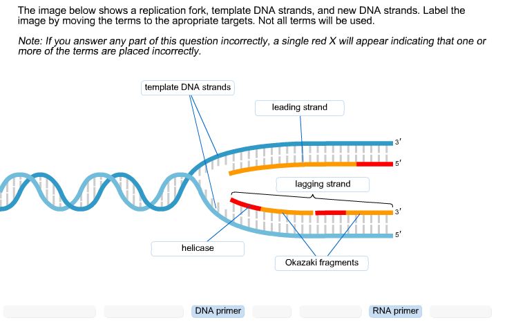
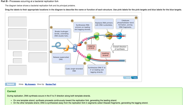
Correctly label the items in this DNA replication fork diagram. Drag the appropriate labels to their respective targets. Labels can be used more than once. Reset | Help Supercoiled chromosome Leading strand DNA polymerase III Lagging strand IIIIIIIIIIIIIIIIIIIIIIIIIIIII Pocong 5' Primase Copcodoro RNA primer Single-strand binding protein Helicase 3

Correctly label the items in this dna replication fork diagram.
DNA: The Molecule Worksheet Spring 2016 ! DNAStructure$ $ 1. On the diagram to the right: • Circle and label a nucleotide. • Place an "s" in the sugar molecules. • Place a "p" in the phosphate molecules. • Label the bases that are not already labeled. • Label a base pair. • Label the sugar-phosphate backbones. 5) DNA ligase links fragments A and B. Synthesis of the lagging strand is accomplished through the repetition of the following steps. Step 1: A new fragment begins with DNA polymerase III binding to the 3' end of the most recently produced RNA primer, primer B in this case, which is closest to the replication fork. In 1958, Meselson and Stahl conducted an experiment to determine which of the three proposed methods of DNA replication was correct. Identify the three proposed models for DNA replication. The Meselson and Stahl experiment starts with E. coli containing 15N/15N15N/15N labeled DNA grown in 14N14N media.
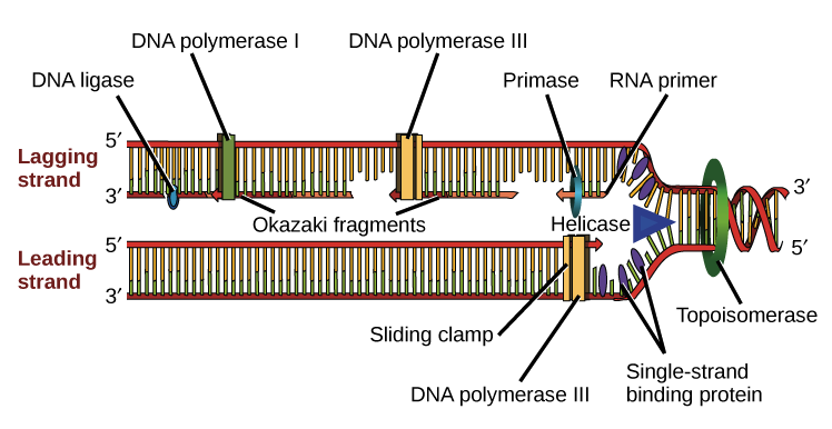
Correctly label the items in this dna replication fork diagram.. The elucidation of the structure of the double helix by James Watson and Francis Crick in 1953 provided a hint as to how DNA is copied during the process of DNA replication.Separating the strands of the double helix would provide two templates for the synthesis of new complementary strands, but exactly how new DNA molecules were constructed was still unclear. An enzyme that unwinds the DNA double helix during DNA replication Okazaki Fragment short segment of DNA synthesized discontinuously in small segments in the 3' to 5' direction by DNA polymerase Use the components in the list below to label the diagram of a replication fork in Figure 2. A. DNA polymerase B. single-strand binding protein C. Okazaki fragment D. primase E. sliding clamp F. RNA primer G. DNA helicase... DNA replication is the production of identical DNA helices from a single double-stranded DNA molecule. Each molecule consists of a strand from the original molecule and a newly formed strand. Prior to replication, the DNA uncoils and strands separate. A replication fork is formed which serves as a template for replication.
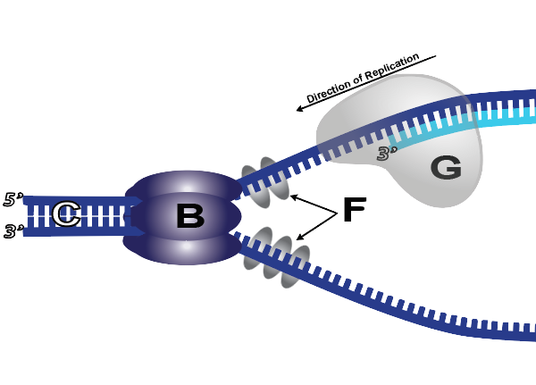
1. Below is a diagram of a DNA replication fork in E. coli. Associate each term or molecular machine with the correct label on the diagram. Some terms may be associated with more than one label, be sure to list ALL that apply. posary D F G Sacaco H C 1.3' end of the lagging strand 2. Correctly label the parts of the two-nucleotide nucleic acid depicted. identify structures and molecules involved in translation. label items in DNA replication fork diagram. In all cells a gene encodes for. a protein, tRNA, or rRNA depending on the specific gene. In this model you can orchestrate the DNA replication fork process that occurs in the cells of all living creatures before cells divide. Both mitosis and meiosis rely on this process to make copies of existing DNA before making new cells. In a real cell, the DNA replication begins by unwinding and unzipping of the twisted double helix structure ... Initially, the simplest mechanism of DNA replication seemed to be the continuous growth of both new strands, nucleotide by nucleotide, at the replication fork as it moves from one end of a DNA molecule to the other. But because of the antiparallel orientation of the two DNA strands in the DNA double helix (see Figure 5-2), this mechanism would require one daughter strand to polymerize in the 5 ...
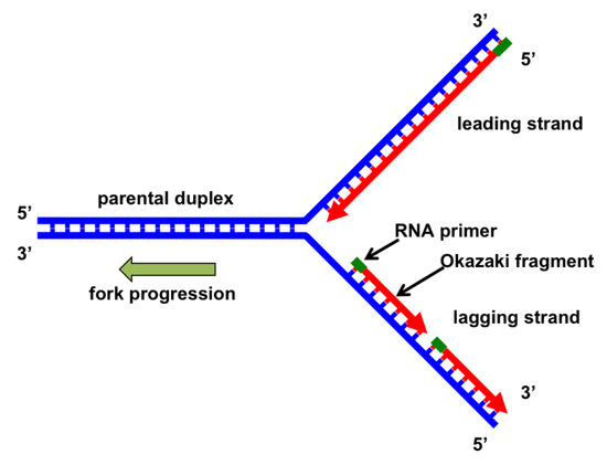
So, DNA replication occurs within a fork-like structure in which the lagging strand loops around in a proposed "trombone model" to orient the replication enzymes for repeated synthesis and joining. It is considered that this mechanism helps to maintain the integrity of DNA during the replication cycles. This quiz is incomplete! To play this quiz, please finish editing it. 6 Questions Show answers. Question 1. SURVEY. 180 seconds. Q. Which letter represents the leading strand? The other two were hybrid molecules (HL). This proves that during replication, one parent strand is conserved and the other new strand is synthesized. Thus DNA replication is a semi-conservative process. Enzymes of DNA Replication: The enzymes which take part in replication are able to copy DNA molecules which may contain millions of bases. 335 draw and label a simple diagram of the molecular structure of dna. Short segment of dna synthesized discontinuously in small segments in the 3 to 5 direction by dna polymerase. Dna replication is discontinuous in one strand. These steps require the use of more than dozen enzymes and protein factors. Here i demonstrate drawing the structure ...
Mastering Biology Exam 3 Flashcards Quizlet
1. Below is a replication fork (one side of an origin): a double-stranded DNA partially opened up to provide single-stranded regions where replication can occur. Draw and label the following: RNA primers (label 3′ and 5′ ends), leading-strand DNA polymerase and new DNA (label 3′ and 5′ ends and show direction of synthesis

Gen Gen Test Uno Flashcards Quizlet
A DNA replication fork looks like a fork in the road and is the place in a DNA molecule where DNA replication occurs. Explore the definition, structure and function, and an overview of a DNA ...
Solved The Image Below Shows A Replication Fork Template Chegg Com
DNA Replication Definition: For the growth of an individual, cell division is a necessary part. When the act of cell division occurs, the DNA must be replicated. During cell division, the DNA successfully copied in the daughter cells. Many enzymes take place for this act. The DNA has to be inherited and copied in two daughter cells.
Genes Free Full Text History Of Dna Helicases Html
Exam 3 Chs 5 Dna Structure And Replication Machinery Amp 16 The Label the parts of the dna replication fork. Label the parts of the dna replication fork. The polymerase has to attach only once and it can continue its work as the replication fork moves forward. Learn vocabulary terms and more with flashcards games and other study tools.
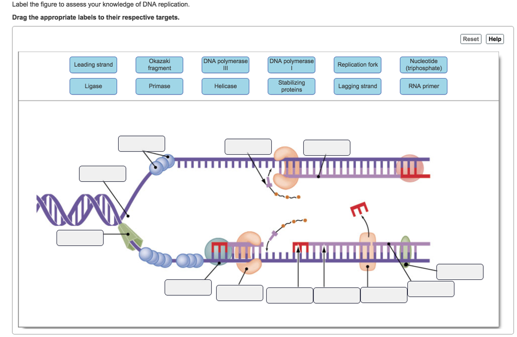
Answered Label The Figure To Assess Your Bartleby
DNA Repair. DNA polymerase can make mistakes while adding nucleotides. It edits the DNA by proofreading every newly added base. Incorrect bases are removed and replaced by the correct base, and then polymerization continues (Figure 9.13 a).Most mistakes are corrected during replication, although when this does not happen, the mismatch repair mechanism is employed.

Cell Cycle Regulation Of Dna Replication Abstract Europe Pmc
Below is a diagram of a DNA replication fork in E. coli. Associate each term or molecular machine with the correct label on the diagram. Some terms may be associated with more than one label, be sure to list ALL that apply. 1.end of the lagging strand 2. Relieves tension due to helicase activity further down the DNA 3.
31 Can You Correctly Label These Images Of Chromosomes Label Design Ideas 2020
Label the diagram: Need help? Check out this Youtube video. SSBs. Label the diagram: DNA polymerase adds nucleotides (5' to 3') Replication fork is formed. DNA polymerase attaches to the primer. Okazaki fragments bound by ligase. ... Stabilizes the DNA molecule during replication ...
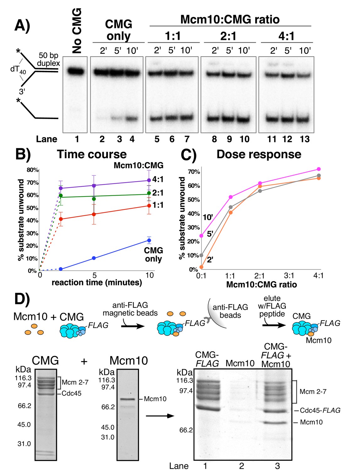
Mcm10 Promotes Rapid Isomerization Of Cmg Dna For Replisome Bypass Of Lagging Strand Dna Blocks Elife
Step 1: A new fragment begins with DNA polymerase III binding to the 3' end of the most recently produced RNA primer, primer B in this case, which is closest to the replication fork. DNA pol III then adds DNA nucleotides in the 5' to 3' direction until it encounters the previous RNA primer, primer A.
1
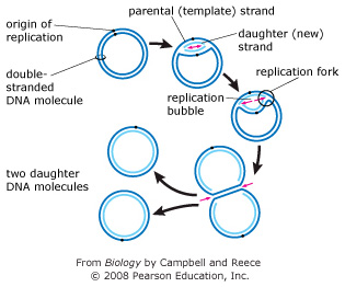
The replication fork is a region where a cell's DNA double helix has been unwound and separated to create an area where DNA polymerases and the other enzymes involved can use each strand as a template to synthesize a new double helix. An enzyme called a helicase catalyzes strand separation. Once the strands are separated, a group of proteins called helper proteins prevent the

Haloferax Volcanii A Model Archaeon For Studying Dna Replication And Repair Open Biology
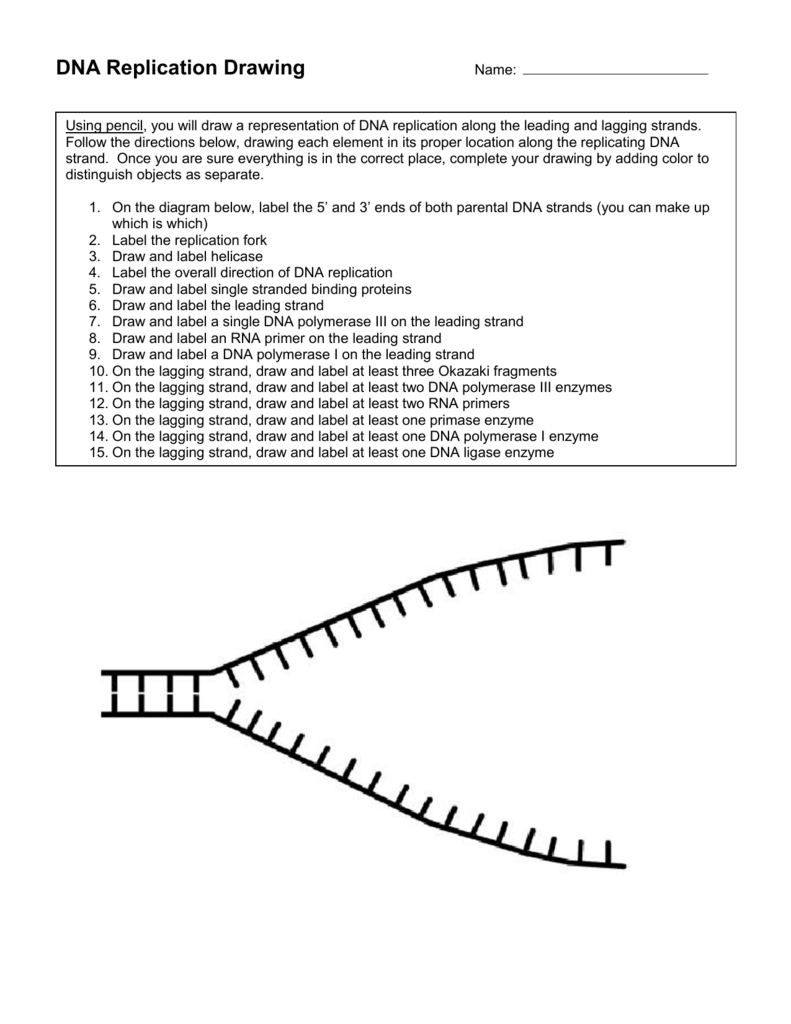
strand. Once you are sure everything is in the correct place, complete your drawing by adding color to distinguish objects as separate. 1. On the diagram below, label the 5' and 3' ends of both parental DNA strands (you can make up which is which) 2. Label the replication fork 3. Draw and label helicase 4.
Dna Replication Fork Drawing
Jun 07, 2020 · Written By Pelvic Diagram Sunday, June 7, 2020. Edit. Dna Replication Diagram Labeled. DNA replication occurs through a semiconservative mechanism, because each new molecule is made up of one old strand and one new strand. DNA Replication has three steps - Initiation, Elongation, and Termination. DNA Replication: Steps, Process, Diagram and ...

Primpol Mediated Adaptive Response Suppresses Replication Fork Reversal In Brca Deficient Cells Sciencedirect
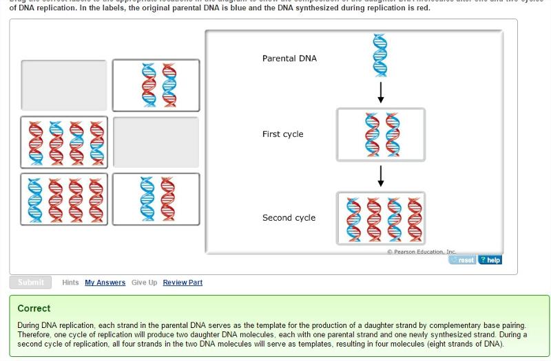
Drag the correct labels to the appropriate locations in the diagram to show the composition of the daughter duplexes after one and two cycles of DNA replication. In the labels, the original parental DNA is blue and the DNA synthesized during replication is red.

Eukaryotic Dna Replication Wikipedia
In this diagram showing the replicationof dna label the following items. Leading and lagging strands okazaki fragment dna polymerase dna ligase helicase primase single stranded binding proteins rna primer replication fork and 5 and 3 ends of the parental dna. The phase diagram for bromine is shown below. The following tabernacle diagrams label ...
Chapter 9 Dna Replication Chemistry
In 1958, Meselson and Stahl conducted an experiment to determine which of the three proposed methods of DNA replication was correct. Identify the three proposed models for DNA replication. The Meselson and Stahl experiment starts with E. coli containing 15N/15N15N/15N labeled DNA grown in 14N14N media.

Dna Replication Microbiology
5) DNA ligase links fragments A and B. Synthesis of the lagging strand is accomplished through the repetition of the following steps. Step 1: A new fragment begins with DNA polymerase III binding to the 3' end of the most recently produced RNA primer, primer B in this case, which is closest to the replication fork.

Ctip Mediated Fork Protection Synergizes With Brca1 To Suppress Genomic Instability Upon Dna Replication Stress Sciencedirect
DNA: The Molecule Worksheet Spring 2016 ! DNAStructure$ $ 1. On the diagram to the right: • Circle and label a nucleotide. • Place an "s" in the sugar molecules. • Place a "p" in the phosphate molecules. • Label the bases that are not already labeled. • Label a base pair. • Label the sugar-phosphate backbones.
Genes Special Issue Dna Replication
Tel Archives Ouvertes Fr
Exam 3 Chs 5 Dna Structure And Replication Machinery 16 The Molecular Basis Of Inheritance Flashcards Easy Notecards
Molecular Mechanism Of Dna Replication Article Khan Academy

Claspin Checkpoint Adaptor And Dna Replication Factor Smits 2019 The Febs Journal Wiley Online Library
Molecules Free Full Text Shining A Spotlight On Dna Single Molecule Methods To Visualise Dna Html
Dna Replication Tutorial Learn Biology

Excessive Excision Of Correct Nucleotides During Dna Synthesis Explained By Replication Hurdles The Embo Journal
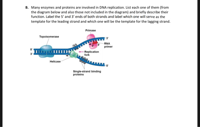
Solved B Many Enzymes And Proteins Are Involved In Dna Chegg Com
Sdjohnston Faculty Noctrl Edu

Chapter 9 Biology 222 Exam 2 Flashcards Quizlet

Biol Chapter 11 Review Flashcards Quizlet

Okazaki Fragments An Overview Sciencedirect Topics

Diagram Of Bacteriophage T4 Proteins And Dna At The Replication Fork Download Scientific Diagram

Pulse Labelling Of Replicating Dna A Dna Replication Starts At The Download Scientific Diagram
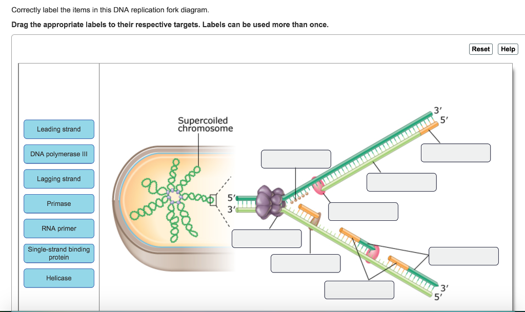
Solved Correctly Label The Items In This Dna Replication Chegg Com
Single Molecule Analysis Reveals That Dna Replication Dynamics Vary Across The Course Of Schizogony In The Malaria Parasite Plasmodium Falciparum Scientific Reports
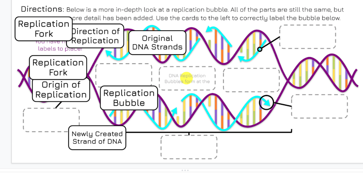
Answered Directions Below Is A More In Depth Bartleby

Eukaryotic Dna Replication Wikipedia
Chapter 16 Molecular Basis Of Inheritance Flashcards Easy Notecards

Mastering Biology Chp 13 Hw Flashcards Quizlet
Mastering Biology Chapter 16 Rhs Homework
Mastering Biology Chapter 16 Rhs Homework
0 Response to "39 correctly label the items in this dna replication fork diagram."
Post a Comment