39 cat muscle anatomy diagram
› books › NBK6229Anatomy and Physiology of the Spinal Cord - Madame Curie ... The spinal cord is part of the central nervous system (CNS), which extends caudally and is protected by the bony structures of the vertebral column. It is covered by the three membranes of the CNS, i.e., the dura mater, arachnoid and the innermost pia mater. In most adult mammals it occupies only the upper two-thirds of the vertebral canal as the growth of the bones composing the vertebral ... External Cat Body Parts Labeled - 15 images - cat external ... [External Cat Body Parts Labeled] - 15 images - dissection of a rat, cat external anatomy illustration science art com, on the cutting edge making connections through cat, fleas by jalees mirza,
en.wikipedia.org › wiki › Shark_anatomyShark anatomy - Wikipedia Rough and rigid placoid scales (dermal denticles) coat the skin of sharks, rays and cartilaginous fishes due to the absence of dermal bone. These scales are present in the dermis, which has fibrous connective tissue components, and project through the epidermis, that contains secretary cells and stratified epidermal cells, to the surface.

Cat muscle anatomy diagram
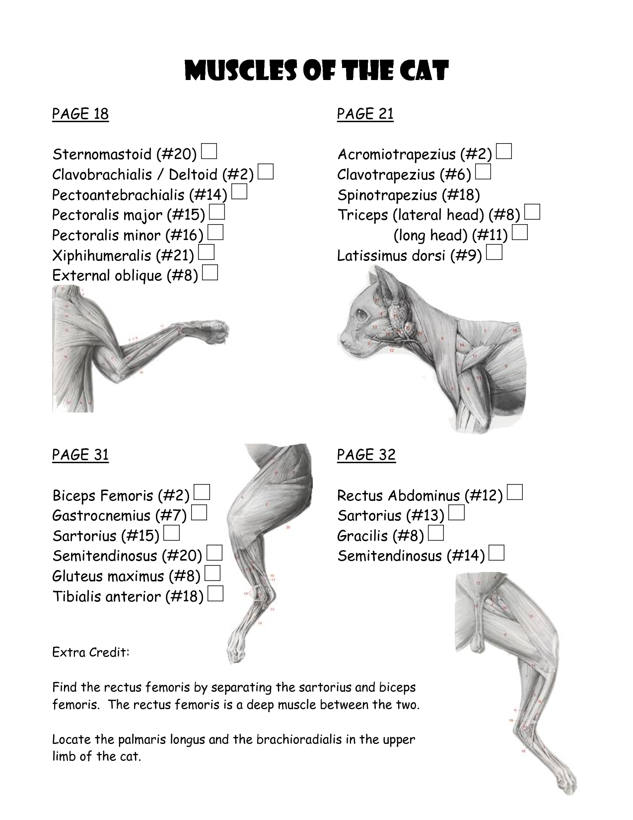
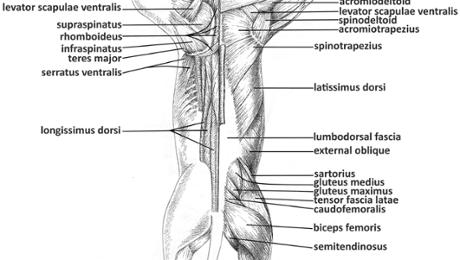
Cat Anatomy Dissection Guide Superficial Muscles. Ventral View pectoantebrachialis. Dorsal View clavotrapezius pectoralis major acromiotrapezius pectoralis minor spinotrapezius.19 pages Chest Wall Muscles Ct Anatomy - anatomy of chest wall and ... Here are a number of highest rated Chest Wall Muscles Ct Anatomy pictures on internet. We identified it from well-behaved source. Its submitted by paperwork in the best field. We undertake this kind of Chest Wall Muscles Ct Anatomy graphic could possibly be the most trending topic subsequent to we allocation it in google improvement or facebook. Large Intestine Anatomy, Parts, Diagram & Major Function ... Anatomy of the Large Intestine. The large intestine is a large organ divided into multiple parts. The large intestine is similar in composition to the small intestine but has a decreased amount of ...
Cat muscle anatomy diagram. Cat Anatomy Labeled - fabulous pets house january 2012 ... Cat Anatomy Labeled - 16 images - anatomy cat stock illustration 536509750 shutterstock, cat anatomy, file scheme cat anatomy wikipedia, domestic cat skull clipart etc, Cat Anatomy and Physiology - Colorado 4-H The cat's muscular control and skeletal flexibility enable it to right its body during a fall with incredible speed—a trick that is unique to cats. Skeleton.44 pages Cat Anatomy Facts For Kids - PoC Cat Anatomy Internal Organs. Diagram from Wikimedia Commons. This is what the organs are: Brain - controls the cat's body. It is the center of the nervous system. Spinal Cord - connects brain to nerves of the body which control the body. Diaphragm - separates lungs from the organs lower down such as the stomach. It helps in breathing. Head and neck anatomy of the cat on CT - vet-Anatomy Head and neck anatomy of the cat on CT - vet-Anatomy Head - Cat - CT (Transversal) Sagittal Dorsal 3D Lateral A subscription is required to unlock all features 1/1152 Revert to the old version of the viewer vet-Anatomy Authors Antoine Micheau - MD , Denis Hoa - MD , Susanne AEB Boroffka - PhD - dipl. ECVDI Published on Monday 13 September 2021
Examining and medicating a cat's ears | Veterinary ... The following picture shows a diagram of the right ear as it appears if you are looking at the cat's head from the front. The outer ear flap is usually covered with fur. If the ear is itchy, scratching may result in hair loss on the ear flap or at the base of the ear. Cat Skeleton Anatomy with Labeled Diagram - AnatomyLearner The radius and ulna of a cat are entirely separated bones in the skeleton. There are seven carpal, five metacarpal, and five digits in the forelimb of cat anatomy. The ischium bone of the cat is twisted, and the boney floor of the pelvis is V-shaped. There is no supracondyloid fossa in the cat's femur bone. Cat Leg Anatomy Diagram - do rabbits have muscles quora ... Cat Leg Anatomy Diagram - 17 images - cat leg bone diagram cat dissection hip thigh and lower, cat dissection lower leg youtube, pin on anatomy, cat anatomy images stock photos vectors shutterstock, byjus.com › biology › facts-about-anatomyAnatomy- Interesting Facts and Information About ... - BYJUS Animal Anatomy. This is also called zootomy. It mainly deals with the study of the internal structure of animals, birds, insects including the cells, tissues, organs, bones and other organs and organs systems of the animal body. Explore more: Anatomy. Let’s know about the amazing and interesting facts about anatomy. Fascinating Facts about ...
kidshealth.org › en › kidsYour Eyes (for Kids) - Nemours KidsHealth A Muscle Makes It Work. The lens is suspended in the eye by a bunch of fibers. These fibers are attached to a muscle called the ciliary (say: SIL-ee-air-ee) body. It has the amazing job of changing the shape of the lens. That's right — the lens actually changes shape right inside your eye! Clitoris: Location, structure, diagram | Kenhub The clitoris is an erectile tissue of the females, located at the junction of the inner lips of vulva and immediately above the external opening of the urethra.The clitoris is responsible for feeling sexual sensations upon stimulation, and in many women, its proper stimulation facilitates orgasm. This article will discuss the anatomy of the clitoris, a female sex organ and most sensitive ... Cat Skull Anatomy with Labeled Diagram » AnatomyLearner ... The cat skull anatomy consists of the cranial and facial bones. These cranial and facial bones of a cat are known as the skull proper. You will find other visceral bones like the lower jaw, hyoid, and ear bone together with the cat skull proper. Here, I will share all the anatomical features of the skull bones from a cat with a diagram. Veterinary Anatomy » AnatomyLearner >> The Place to Learn ... Cat Skull Anatomy with Labeled Diagram 12/03/2022 12/03/2022 by anatomylearner The cat skull anatomy consists of the cranial and facial bones. These cranial and facial bones of a cat are known as the skull proper. You will find other visceral bones like the lower jaw, hyoid, and ear bone together with the cat skull proper.
Cat Dissection - A Supplemental Guide • bluedoor Publishing
CT scan of head and neck - e-Anatomy - IMAIOS CT scan of head and neck : Radiological anatomy of the head and neck on a CT in axial, coronal, and sagittal sections, and on a 3D images. CT scan of head and neck : Axial. CT scan of head and neck : Axial. CT scan of head and neck : Sagittal. CT scan of head and neck : Coronal.

Cat muscle anatomy dorsal Diagram | Quizlet
CT arthrogram of the shoulder joint - e-Anatomy Atlas of the anatomy of the joint of the shoulder on a CT arthrogram in axial, coronal, and sagittal sections, on a 3D images and on conventional athrogram

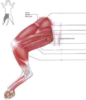
Muscle Human musculoskeletal system Arm Cat anatomy, arm ...
Anatomy Cat Dissection Muscles - cat muscles upper body ... Anatomy Cat Dissection Muscles. Here are a number of highest rated Anatomy Cat Dissection Muscles pictures upon internet. We identified it from honorable source. Its submitted by executive in the best field. We tolerate this nice of Anatomy Cat Dissection Muscles graphic could possibly be the most ...

Human Anatomy & Physiology 1
Scalene muscles: Innervation, function, action, location ... The scalene muscles are the three muscles found on each side of the neck, spanning between the transverse processes of the cervical vertebrae and the upper two ribs. Namely, these muscles are the scalenus anterior (anterior scalene), scalenus medius (middle scalene) and scalenus posterior (posterior scalene).

Masticatory muscles in the dog (left) and cat (right)-lateral ...
› pmc › articlesThe thoracolumbar fascia: anatomy, function and clinical ... May 27, 2012 · The TrA muscle, based on its anatomy and function, has the greatest potential for influencing the stability of the lumbar spine (Hodges et al. 2003; Barker et al. 2007). Its role in respiratory movement has been reviewed by De Troyer et al. (1990). De Troyer (2005), and its role in postural stability has been reviewed by Hodges (1999, 2008 ...

Thumb Muscle Head and neck anatomy Head and neck anatomy ...
Learn all muscles with quizzes and labeled diagrams | Kenhub Human body muscle diagrams. Muscle diagrams are a great way to get an overview of all of the muscles within a body region. Studying these is an ideal first step before moving onto the more advanced practices of muscle labeling and quizzes. If you're looking for a speedy way to learn muscle anatomy, look no further than our anatomy crash courses .
cat muscle work template : Biological Science Picture ...
byjus.com › biology › thyroid-glandThyroid Gland - Anatomy, Location, Function, Disorders Thyroid Gland is a butterfly-shaped endocrine gland situated in the anterior portion of the neck. It is responsible for the production of thyroid hormones that affect important functions in the body

3D Cat Anatomy
anatomylearner.com › cow-anatomyCow Anatomy - External Body Parts and Internal Organs with ... You will find a shallow musculosprial groove for the brachialis muscles in the humerus of a cow. The deltoid tuberosity of cow is less prominent than in horses. You will find a large greater tubercle that forms the point of the shoulder in a cow. The radius of a cow is relatively short but broad than those of a horse.

Cheetah Anatomy Chart
Cat and dog anatomy | Veterinary Teaching Hospital ... Cat Lymphatic System The Lymphatic System includes lymph nodes and lymph vessels throughout the body. Musculoskeletal System The musculoskeletal system includes all the muscles, bones and joints. Shoulder Elbow Rear Leg Stifle (knee) Shoulder Elbow Rear Leg Stifle (knee) Respiratory System

Way I understand the world: Interesting Facts about Cats I ...
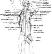
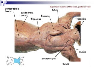
Anatomy & Physiology Cat Muscles – Ventral view 1 - Moore ... Anatomy & Physiology. Cat Muscles – Ventral view 1. Page 2. Anatomy & Physiology. Cat Muscles– Ventral view 2 ... Cat Muscles – Lateral view 1 ...9 pages
Cat Muscle Dissection--Dorsal Muscles of Back, Arm, Shoulder ...
Basic Horse Anatomy: Part 1 - The Open Sanctuary Project There are several diagrams in this resource covering basic body anatomy and basic hoof anatomy. After each diagram, there will be a glossary of terms used in the diagram to provide more clarity. Let's start out looking at a diagram showing basic horse anatomy. Knowing the vocabulary and the areas they refer to on a horse resident's body ...

Felidae Cat anatomy Cat anatomy Organ, Cat, animals, anatomy ...
Cat Anatomy Organs - 17 images - cat anatomy domestic cats ... [Cat Anatomy Organs] - 17 images - pancreas l jpg, feline anatomy 101 the conscious cat, links to pictures on the physiology of cats, biology cat digestive tract youtube,

Cat Muscles – PoC
Levels Of Cat Muscle Quiz - ProProfs Try this amazing Levels Of Cat Muscle Quiz quiz which has been attempted 3271 times by avid quiz takers. Also explore over 102 similar quizzes in this category.
Comparative Anatomy - Cat Muscles Flashcards | Chegg.com
Knee Joint Anatomy Diagram Muscle - anatomy of the knee ... Here are a number of highest rated Knee Joint Anatomy Diagram Muscle pictures upon internet. We identified it from obedient source. Its submitted by meting out in the best field. We allow this nice of Knee Joint Anatomy Diagram Muscle graphic could possibly be the most trending subject considering we portion it in google gain or facebook.
anatomy I: practical 3: Cat Muscles Flashcards | Chegg.com
Neck Anatomy: Muscles, glands, organs | Kenhub Neck spaces. The content of the neck is grouped into 4 neck spaces, called the compartments.. Vertebral compartment: contains cervical vertebrae and postural muscles.; Visceral compartment: contains glands (thyroid, parathyroid, and thymus), the larynx, pharynx and trachea.; Two vascular compartments: contain the common carotid artery, internal jugular vein and the vagus nerve, on each side of ...

Diagram of innervation of pharynx in cat. A, artery; M ...
Cat Skeletal Anatomy - Exploring Nature Cat Skeletal Anatomy, cat skeleton. When you research information you must cite the reference. Citing for websites is different from citing from books, magazines and periodicals.
Dissected Quotes. QuotesGram
Cat Paw Anatomy - Bone, Muscle, and Digital Pad ... The most important muscles of the cat front paws are extensor digitorum communis, extensor digitorum lateralis, flexor digitorum profundus, flexor digitorum superficialis, and lumbrical muscles. These muscles form the strong ligaments and tendons, important structures in the cat's front paw.

Levels Of Cat Muscle Quiz - ProProfs Quiz
Anatomy Coloring Book - blank ear diagram human ear ... Anatomy Coloring Book. Here are a number of highest rated Anatomy Coloring Book pictures upon internet. We identified it from obedient source. Its submitted by meting out in the best field. We believe this kind of Anatomy Coloring Book graphic could possibly be the most trending subject bearing in mind we allowance it in google help or facebook.

Anatomy - Unit 5 (Cat Muscles) Diagram | Quizlet
10 Dog Anatomy Worksheets in 2022 ᐅ The Poetry House By using worksheets you have ...
Cat Muscles Lab Guide
Abdomen and pelvis anatomy of the dog on CT - vet-Anatomy CT images are from a healthy 6-year-old castrated male dog. In this module of the animal atlas vet-Anatomy is displayed the cross-sectional labeled anatomy of the canine abdominal cavity and the pelvis on a Computed Tomography (CT) and on 3D images of the abdomen of the dog. CT images are available in 3 different planes (transverse, sagittal ...
Cat Anatomy Dissection Guide
Large Intestine Anatomy, Parts, Diagram & Major Function ... Anatomy of the Large Intestine. The large intestine is a large organ divided into multiple parts. The large intestine is similar in composition to the small intestine but has a decreased amount of ...

Cat muscles | Cat anatomy, Anatomy, Vet medicine
Chest Wall Muscles Ct Anatomy - anatomy of chest wall and ... Here are a number of highest rated Chest Wall Muscles Ct Anatomy pictures on internet. We identified it from well-behaved source. Its submitted by paperwork in the best field. We undertake this kind of Chest Wall Muscles Ct Anatomy graphic could possibly be the most trending topic subsequent to we allocation it in google improvement or facebook.
Cat Muscle Anatomy with Labeled Diagram » AnatomyLearner ...
Cat Anatomy Dissection Guide Superficial Muscles. Ventral View pectoantebrachialis. Dorsal View clavotrapezius pectoralis major acromiotrapezius pectoralis minor spinotrapezius.19 pages

Kennedy, Anne / Anatomy Review

Cat musculature | Atlas of Comparative Vertebrate Anatomy

Cat musculature | Atlas of Comparative Vertebrate Anatomy

cat muscles Diagram | Quizlet
Cat Muscle Dissection--Muscles of Chest, Neck, Arms, and ...

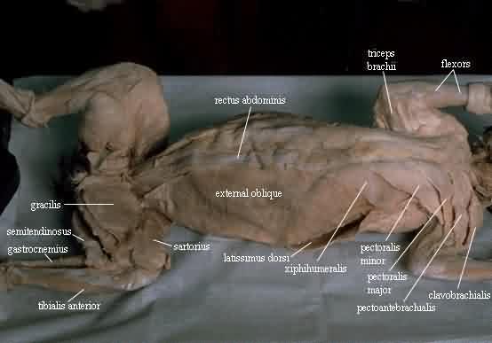
Pectoralis Muscles of the Cat

Cat Muscles of the Chest (color)
Cat dissection lab_labeled_images

Munchkin Cat Muscle Anatomy by TheDragonofDoom on DeviantArt

Cat musculature | Atlas of Comparative Vertebrate Anatomy
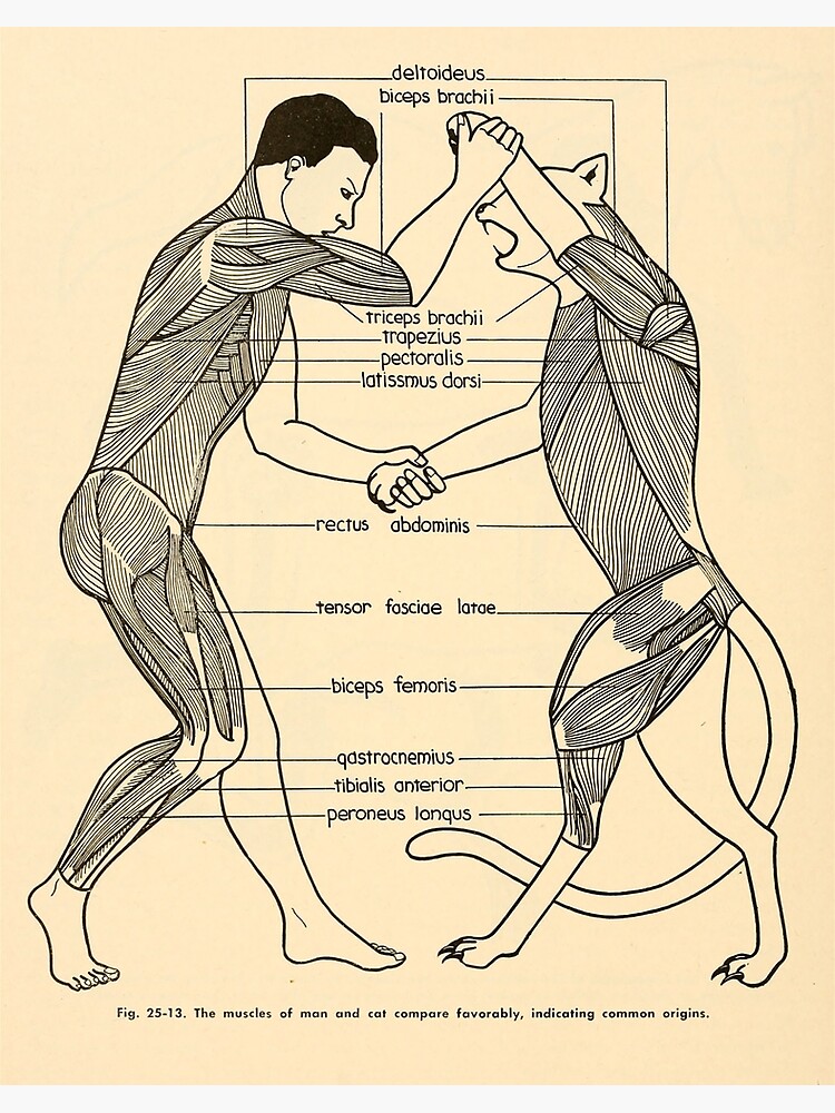
Evolution of Cat vs Human Anatomy Diagram" Art Board Print by ...
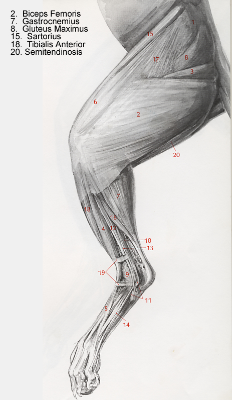
Identify the numbered muscles of the cat in figures 62.25 ...

Cat anatomy Finger Muscle Neck, Cat, animals, hand, human png ...
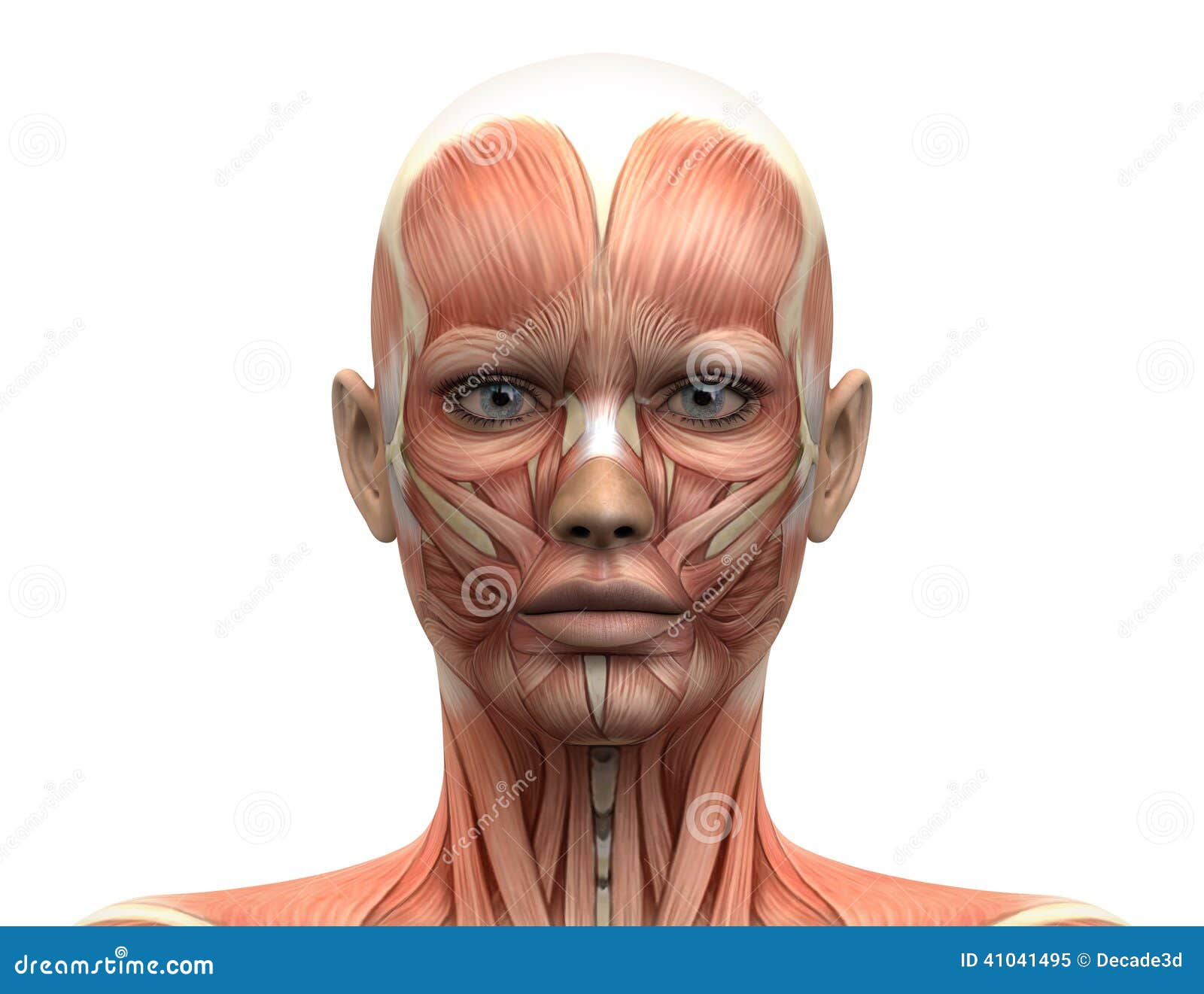
Female Head Muscles Anatomy - Front View Stock Illustration ...
Anatomy & Physiology Cat Muscles – Ventral view 1

Cat anatomy - Wikipedia
0 Response to "39 cat muscle anatomy diagram"
Post a Comment