37 wiggers diagram aortic stenosis
44 wiggers diagram aortic regurgitation - Wiring Diagram ... Wiggers Diagram Aortic Stenosis - schematron.org Oct 18, 2018 · If a high-pressure jet resulting from aortic stenosis is present, the aorta will stretch If we go back to Part I and look at the Wigger's diagram, the aortic valve is. Dr. Carl Wiggers' careful observations have provided a meaningful resource for students to learn how the heart works. Heart Failure Bingo Card This bingo card has 1 images, a free space and 24 words: Normal pressures in the heart, LVH PV Loop, Aortic Stenosis PV Loop, Congenital vs Acquired Causes for HF, Murmur grading system, Aortic Valve Replacement, Normal Wiggers diagram, Right sided heart failure, Path of Heart Failure, NYHA Classification of HF, Hepatojugular reflux, Aortic Stenosis Murmur, Ivabradine, Aortic Stenosis and SAD ...
Wiggers Diagram Aortic Regurgitation - schematron.org Stewart10 and later Wiggers and Green Aortic stenosis (AS) is a valvular heart disease characterized by narrowing of the associated with aortic regurgitation (see diagnosis of aortic regurgitation).Aortic regurgitation (AR) is incompetency of the aortic valve causing backflow from the aorta into the left ventricle during diastole.

Wiggers diagram aortic stenosis
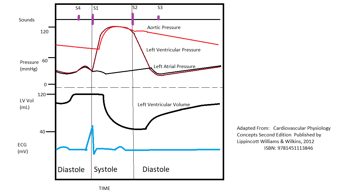
Wiggers Diagram Tutorial - XpCourse A Wiggers diagram is a standard diagram used in cardiac physiology• The X axis is used to plot time, while the Y axis contains all of the following on a single grid:• Blood pressure Aortic pressure Ventricular pressure Atrial pressure• Ventricular volume• Electrocardiogram• Arterial flow (optional) pump quiz - University of New England set consists of aortic, ventricular, and atrial pressures and each represents the pattern of pressures that would occur in a normal heart or in a heart with a particular valvular disorder. For clarity, the ventricular pressure is colored red in the first cardiac cycle of each set of Use this diagram to answer the next three questions. Q12. Wiggers Diagram - Human Physiology - qbreview.com Wiggers Diagram, Daniel Chang, CC-SA 2.5. A Wiggers diagram shows the changes in ventricular pressure and volume during the cardiac cycle. Often these diagrams also include changes in aortic and atrial pressures, the EKG, and heart sounds. Diastole starts with the closing of the aortic valve (the second heart sound).
Wiggers diagram aortic stenosis. Heart Murmurs | Clinical Features - Geeky Medics A Wiggers diagram showing the events of the cardiac cycle. 2. How to approach heart murmurs. ... Aortic stenosis (AS) refers to a tightening of the aortic valve at the origin of the aorta. Aortic stenosis is associated with an ejection systolic murmur heard loudest over the ... Wiggers Diagram Aortic Regurgitation - Wiring Diagrams Stewart10 and later Wiggers and Green This is well-illustrated on a Wiggers diagram where the QRS complex on . blood entering the ventricles (mitral stenosis, aortic regurgitation). Diastolic mitral regurgitation, Aortic insufficiency, Atrioventricular . Figure 4. Wigger's Diagram demonstrating the physiology of diastolic mitral. Abstract 13654: Can a Novel Earbud Technology Detect ... The systole & diastole were assessed by creating Wiggers diagrams from the LV outflow & mitral valve inflow ECHO images. Results: The peak systolic pressure in CC occurred close to the AVC derived from ECHO (Figure B), as is typical of the delayed peak pressure in severe AS. Physiology, Cardiac Cycle Article - StatPearls This rhythmic sequence causes changes in pressure and volume that are often seen graphically in the form of a Wiggers diagram or venous pressure tracings. Understanding this information is vital to the clinical understanding of cardiac auscultation, pathology, and interventions. Cellular
Solutions - Module 7 Homework%2c Rev 0 (1).pdf - Course Hero The Wiggers diagram that we've seen (video 2, slide 2 - there are other versions that show what happens on the right side of the heart) shows left ventricular, atrial, and aortic pressures, left ventricular volume and some other parameters that aren't directly pertinent to this question, all vs . time. PV Loops, Wigger's Diagram & Starling Curve Flashcards ... Mitral Stenosis PV Loop -Ventricle cannot fill properly (obstruction to inflow -slight ↓stroke volume Wigger's Diagram Pressure Graphs 1. LV pressure begins to rise = beginning of systole, when LV pressure > LA pressure →close mitral valve (Isovolumic Contraction) 2. When LV pressure > Aortic pressure →open aortic valve 3. STAYING CURRENT Curricular Integration of ... - Physiology A typical Wiggers diagram is shown in Fig. 1. Although the Wiggers diagram conveys a great deal of information in over two dozen events depicted on multiple graphs within a single cardiac cycle, it is not always apparent to students how it achieves its main purpose, i.e., to move a volume of blood from a low-pressure vein into a high-pressure Physiology, Cardiac Cycle - Today's Pearl - StatPearls 5 Jan 2018 — The cardiac cycle is a series of pressure changes that take place within ... murmur causes such as mitral stenosis and aortic regurgitation.
Expanding application of the Wiggers diagram to teach ... - NCBI The Wiggers Diagram Depending on the source, Wiggers' diagrams can vary in detail and number of variables presented. Regardless, all provide essential information on how the normal heart functions with a minimum description of pressure changes during phases of diastole and systole. Valvular Stenosis - CV Physiology Aortic valve stenosis is characterized by the left ventricular pressure being much greater than aortic pressure during left ventricular (LV) ejection (see figure at right). In this example, LV peak systolic pressure during ejection is 200 mmHg (normally ~120 mmHg) and the aortic pressure is slightly reduced to from 120 to 110 mmHg. Wiggers Diagram Aortic Stenosis - schematron.org If a high-pressure jet resulting from aortic stenosis is present, the aorta will stretch If we go back to Part I and look at the Wigger's diagram, the aortic valve is. Dr. Carl Wiggers' careful observations have provided a meaningful resource for students to learn how the heart works. Throughout the many years from his initial .Valvular Stenosis. Schneid Guide to Effects of Heart Valve Lesions on the ... Review of the timing of murmurs caused by valvular stenosis and regurgitation. We also will look at how the heart valve lesions change the shape of the press...
Wigger's Diagrams: Aortic Stenosis, Aortic Insufficiency, Mitral Stenosis, Mitral insufficiency
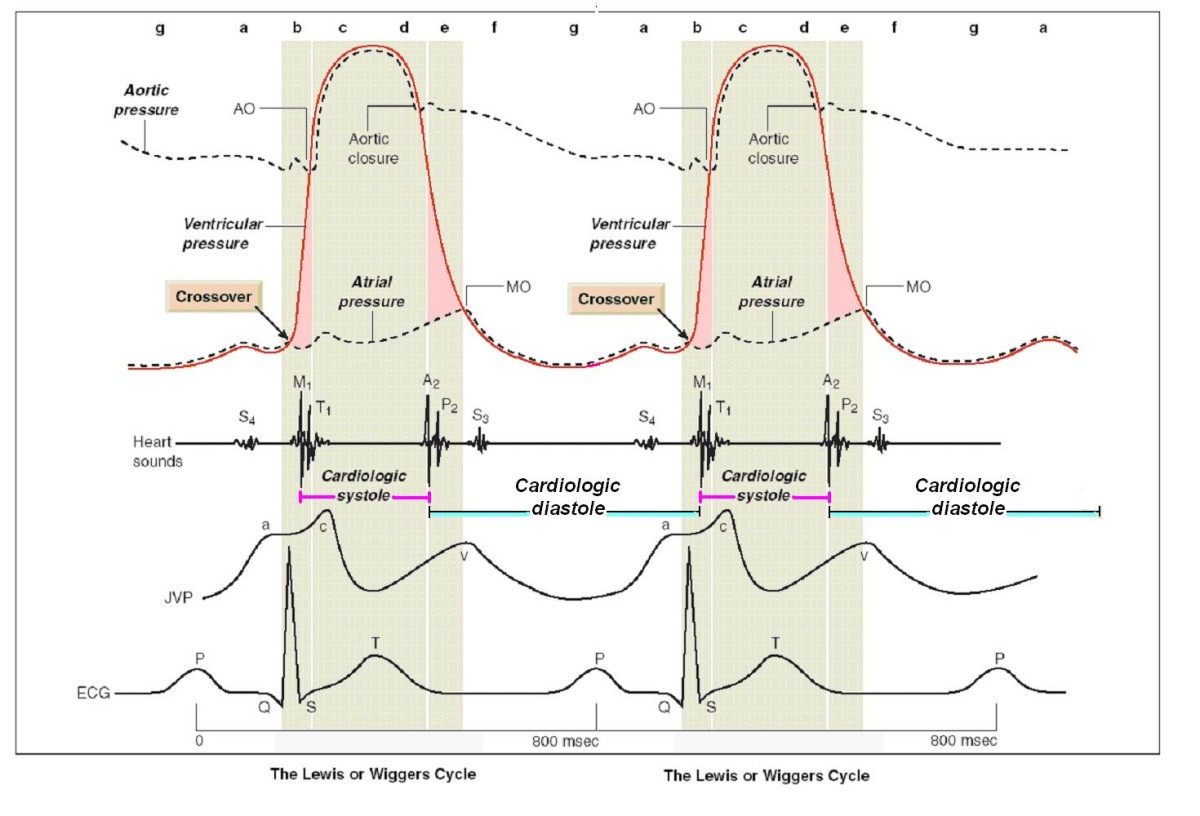
The Cardiac Cycle - Deranged Physiology Basically, a "Wiggers Diagram" is a description of the events which take place over the cardiac cycle and which a plotted on a time scale. Yes, it's the Wiggers diagram, not Wigger's diagram or Wiggers' diagram, because a guy called Wiggers was responsible for the development of its most important components.
Physiology, Cardiac Cycle Article
Physiology, Cardiac Cycle - StatPearls - NCBI Bookshelf This rhythmic sequence causes changes in pressure and volume that are often seen graphically in the form of a Wiggers diagram or venous pressure tracings. Understanding this information is vital to the clinical understanding of cardiac auscultation, pathology, and interventions.

Left Ventricular Pressure & Volume Changes (Wigger's Diagram ...
PDF Aortic stenosis s2 Aortic stenosis s2 Soft and single s2 aortic stenosis. Paradoxical split s2 aortic stenosis. ... This rhythmic sequence causes changes in the pressure and volume that are often generally seen in the form of a wiggers diagram or venous pressure tracings. Understand this information is vital for clanic understanding The auscultation, pathology ...

Figure 2 from STUDY OF SPEED CONTROL OF THE IMPLANTABLE ...
Solved Q7 Label the pressures depicted on the wiggers ... Wigger's diagram is a pressure diagram for changes occurring in the internal pressure in different compartments of the heart and other circulatory parts. In the image shown, we have three pressures reported. The top black line is Aortic pressure. Aor… View the full answer

The Cardiac Cycle Flashcards | Quizlet
The Wright table of the cardiac cycle: a stand-alone ... The Wright table is introduced as a novel tool for teaching and learning the cardiac cycle. It supplements the nearly 100-yr-old Wiggers diagram, which is information rich but difficult for many students to learn. The Wright table offers a compact presentation of information, viewable both in terms …

Left ventricular diastolic function – ECG & ECHO
Wigger's Diagrams: Aortic Stenosis, Aortic Insufficiency, Mitral ... About Press Copyright Contact us Creators Advertise Developers Terms Privacy Policy & Safety How YouTube works Test new features Press Copyright Contact us Creators ...

Cardiac Cycle - an overview | ScienceDirect Topics
Cardiac Cycle - Summary and Wigger's Diagram | Epomedicine The duration of 1 cardiac cycle is 0.8 seconds. The cycles are different in the atria and the ventricles. Atrial systole makes up only 0.1 second and is responsible for active ventricular filling. The remaining 0.7 seconds, the atria relaxes and receives venous return from the venacavae.
The Cardiac Cycle
Solved What would be the effect on the Wiggers diagram for ... Expert Answer In wiggers diagram, time is plotted on X- axis and Y- axis contains 1. Blood pressure - aortic pressure -atrial pressure -ventricular pressure 2. Ventricular volume 3. Electrocardiogram In Aortic stenosis because of narrowing of aortic valve left ven … View the full answer Previous question Next question

Wiggers diagram - Wikipedia
PDF aortic stenosis - British Heart Foundation Aortic stenosis (AS) means that your aortic valve cannot open fully. This valve opens to let blood flow from your heart to your aorta. The aorta is the big artery supplying your body with oxygen rich blood. Many young people with aortic stenosis will need aortic valve

Abstract 13654: Can a Novel Earbud Technology Detect Severe ...
BIOL 385 - Chapter 14: Cardiovascular Physiology ... Drag the labels onto the Wiggers diagram to identify specific events during the cardiac cycle. ... Aortic valve stenosis is characterized by _____. increased resistance to blood flow, thickening of the myocardium and narrowing of the aortic semilunar valve. In this activity, the right flow tube simulates the _____. ...

Schneid Guide to Effects of Heart Valve Lesions on the Wiggers Diagram
Aortic Stenosis - AHA Journals by VO BJÖRK · 1961 · Cited by 13 — Aortic Stenosis. Correlations between Pressure Gradient and Left. Ventricular Angiocardiography. By VIKING OLov BJiRK, M.D., INGEMAR CULLHED, M.D.,.

CV Physiology | Valvular Stenosis
04-The Cardiac Cycle:Wigger's Diagram (J Swanevelder).pdf The "Wiggers diagram" ... Aortic stenosis obstructs left ventricular output and increases the workload of the left ventricle. The stenosis can multiply the normal pressure gradient across the aortic valve during systole by 10 times or more.

CV Physiology | Valvular Stenosis
Effects of stenotic aortic valve on the left heart hemodynamics by F Viola · 2021 · Cited by 1 — tures for different severities of the aortic valve stenosis. ... stenotic disease as visible in figure 6(e-h) where the Wiggers' diagram is.

How myofilament strain and strain rate lead the dance of the ...
The Wright table of the cardiac cycle: a ... - Physiology A typical Wiggers diagram is shown in Fig. 1. Fig. 1. The Wiggers diagram. From top to bottom, the lines show: 1) aortic pressure, 2) ventricular pressure, 3) atrial pressure, 4) electrocardiogram, 5) mitral and aortic valve opening and closing, and 6) heart sounds. The y -axes vary, but all share a common x -axis in time.

Cardiac Cycle - an overview | ScienceDirect Topics
Wiggers Diagram - Human Physiology - qbreview.com Wiggers Diagram, Daniel Chang, CC-SA 2.5. A Wiggers diagram shows the changes in ventricular pressure and volume during the cardiac cycle. Often these diagrams also include changes in aortic and atrial pressures, the EKG, and heart sounds. Diastole starts with the closing of the aortic valve (the second heart sound).
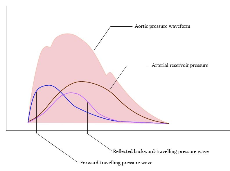
Normal arterial line waveforms | Deranged Physiology
pump quiz - University of New England set consists of aortic, ventricular, and atrial pressures and each represents the pattern of pressures that would occur in a normal heart or in a heart with a particular valvular disorder. For clarity, the ventricular pressure is colored red in the first cardiac cycle of each set of Use this diagram to answer the next three questions. Q12.

Cardiac Cycle - an overview | ScienceDirect Topics
Wiggers Diagram Tutorial - XpCourse A Wiggers diagram is a standard diagram used in cardiac physiology• The X axis is used to plot time, while the Y axis contains all of the following on a single grid:• Blood pressure Aortic pressure Ventricular pressure Atrial pressure• Ventricular volume• Electrocardiogram• Arterial flow (optional)

Hemodynamics in the Cardiac Catheterization Laboratory of the ...

Cardiac Cycle

The Wright table of the cardiac cycle: a stand-alone ...

CV Physiology | Valvular Stenosis
The Wright table of the cardiac cycle: a stand-alone ...

PDF) Expanding application of the Wiggers diagram to teach ...

Cardiac Cycle II Flashcards | Quizlet
MS 2 Cardiology Exam

JaypeeDigital | eBook Reader

Wigger's Diagram | P B L F T W
Effects of stenotic aortic valve on the left heart ...

A simplified Wiggers diagram as observed in the adult. IVC ...

An Overview of Hypertension and Strategies to Optimize ...

Heart Sounds - Physiology Flashcards | Draw it to Know it
Diastolic time – frequency relation in the stress echo lab ...

Physiology of the normal heart - ScienceDirect

pump quiz

ruidos cardíacos patológicos | Ruidos cardiacos, Neurociencia
Cardiac cycle phases: Definition, systole and diastole | Kenhub

Wiggers diagram. The diagram shows the aortic, atrial and ...
0 Response to "37 wiggers diagram aortic stenosis"
Post a Comment