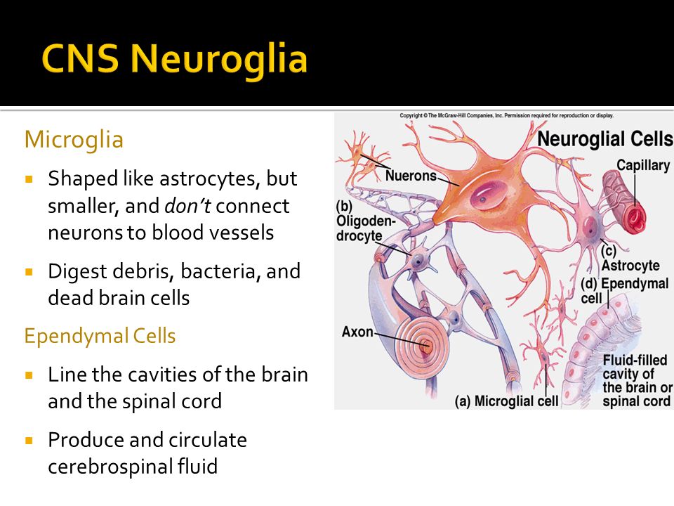
37 identify which diagram represents cells that produce and circulate cerebrospinal fluid.
cerebrospinal fluid is secreted by the quizlet The ventricles are structures that produce cerebrospinal fluid, and transport it around the cranial cavity.They are lined by ependymal cells, which form a structure called the choroid plexus.It is within the choroid plexus that CSF is produced.. Embryologically, the ventricular system is derived from the lumen of the neural tube. Anatomy & Physiology - Anatomy And Physiology, Helping ... The Sertoli cells produce androgen-binding protein. D. The seminal fluid is alkaline and rich in nutrients. ... The glial cell that helps to circulate cerebrospinal fluid is the. A. astrocyte. B. ependymal cell. C. neurolemmocyte. D. microglial cell. ... _____ Produces 60% of the seminal fluid and supplies the sperm with fructose
2injectionsdiabetes 💯compared to type 1 Aug 26, 2021 · The implications for our results were assessed by the fail-safe N and the trim-and-fill method .|A flow diagram of search and selection is shown in Figure 1. The database search resulted in 6258 (6039 Pubmed, 117 Lilacs, 102 Cochrane Trials) articles and 29 other articles were identified from the references of included studies, totalizing 6287 ...
Identify which diagram represents cells that produce and circulate cerebrospinal fluid.
Identify which diagram represents cells that produce and ... Identify which diagram represents cells that produce and circulate cerebrospinal from BIO 005 at Sierra College. Study Resources. Main Menu; ... controlling ion levels in the environment around neurons help with synapse formation in the developing nervous system produce cerebrospinal fluid. 29 Identify Which Diagram Represents Cells That Produce ... Interneurons are an embryonic cell type that becomes a neuron. Which of the labeled cells in the diagram is a neuroglial cell that produces and assists in the circulation of cerebrospinal fluid. Start studying chapter 12. Interneurons are unipolar neurons whose cell bodies are located in the dorsal root ganglia. Neurons and Glial Cells | Boundless Biology Ependymal cells line fluid-filled ventricles of the brain and central canal of the spinal cord which produce cerebrospinal fluid. Key Terms. satellite glia: glial cell that provides nutrients for neurons in the PNS; radial glia: glial cell that serves as a bridge for developing neurons as they move to their end destinations
Identify which diagram represents cells that produce and circulate cerebrospinal fluid.. Anatomy Final Flashcards - Quizlet Identify which diagram represents cells that produce and circulate cerebrospinal fluid. B. Identify the letter that indicates the region of a neuron with a name that means "little hill." C. Identify which diagram represents a microglial cell. E. Identify which letter represents an oligodendrocyte. A. Free Anatomy Flashcards about KATHLEEN MARAVILLAS The regions of the neuron direct electrical currents toward the cell body. DENDRITE: Identify which diagram represents cells that produce and circulate cerebrospinal fluid. D - CEREBROSPINAL FLUID: The somatic nervous system carries information to the: SKELETAL MUSCLES: The ____ of a presynaptic neuron associates with the dendrite of a ... (PDF) Goodman & Gilman’s The Pharmacological Basis of ... Academia.edu is a platform for academics to share research papers. Free Anatomy Flashcards about Anatomy Before Final Identify which diagram represents cells that produce and circulate cerebrospinal fluid. D: The efferent pathways of the autonomic nervous system can be divided into the _____ divisions. parasympathetic and sympathetic : Neurons in the CNS have less chance of regenerating for all of the following reasons except: microglia lay down scar tissue.
Cerebrospinal fluid flow: Anatomy and functions | Kenhub Circulation Cerebrospinal fluid is constantly produced at a secretion rate of 0.2-0.7 ml/min, meaning that there is 600-700 ml of newly produced CSF per day. Since the total volume of CSF averages around 150-270 mL, this means that the entire volume of CSF is replaced around 4 times per day. The pathway of the cerebrospinal fluid is as follows: nervous system . summer class questions part 1 Flashcards ... A. Identify which diagram represents a microglial cell. Identify which diagram represents a cell that produces a myelin sheath in the central nervous system. Identify which letter represents an oligodendrocyte. Identify which diagram represents cells that produce and circulate cerebrospinal fluid. Week 8 Homework_ SCI220-01 Human Anatomy ... - Course Hero 1 / 1 pts Question 15 Use the diagrams above to answer the following question. 10/24/2019 Week 8 Homework: SCI220-01 Human Anatomy with Lab 2019-3 9/9 Identify which of the diagrams represents cells that produce and circulate cerebrospinal fluid. Free Biology Flashcards about Anatomy Identify which diagram represents cells that produce and circulate cerebrospinal fluid. D: Identify the letter that indicates the region of a neuron where neurotransmitters are released. E: This tends to be the longest cytoplasmic projection from a neuron. AXON: Identify the letter that indicates a Schwann cell. C: Bundles of myelinated fibers ...
Free Flashcards about 6-12 Identify which diagram represents cells that produce and circulate cerebrospinal fluid. D : Chemical signals diffuse between neurons at this location. axon terminal : The _____ nervous system is a complex network of nerve pathways embedded in the intestinal wall with a network of integrators and feedback loops that can act somewhat ... Free Flashcards about Stack #3356819 Stack #3356819. Question. Answer. Identify which diagram represents a cell that produces a myelin sheath in the central nervous system. E. Identify the letter that indicates a Schwann cell. C. Identify which diagram represents cells that produce and circulate cerebrospinal fluid. (PDF) Carbohydrates - ResearchGate Living cells produce only one chiral form of biomolecules because the enzymes that synthesize them are also chiral. Stereospecificity, the ability to distinguish betw een stereoisomers, is a ... Join LiveJournal Password requirements: 6 to 30 characters long; ASCII characters only (characters found on a standard US keyboard); must contain at least 4 different symbols;
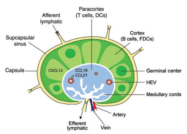
Lymphoid organ development: from ontogeny to neogenesis ...
Week 7_ Activity_ SCI220-16 Human Anatomy with Lab 2021-3 ... Correct! axon terminal axon cell body dendrite 1 / 1 pts Question 17 Figure 12.1 Use the diagram pictured above to answer the following questions. 10/12/21, 3:04 PM Week 7: Activity: SCI220-16 Human Anatomy with Lab 2021-3 11/19 Identify the letter that indicates the gaps between Schwann cells that are known as myelin sheath gaps (nodes of ...

Dementia-associated changes of immune cell composition within ...
Ch. 12 Nervous Tissue Diagram | Quizlet Identify which diagram represents a cell that produces a myelin sheath in the central nervous system. Identify which letter represents an oligodendrocyte. Identify which diagram represents cells that produce and circulate cerebrospinal fluid. A neuron is a collection of nerve fibers in the PNS.
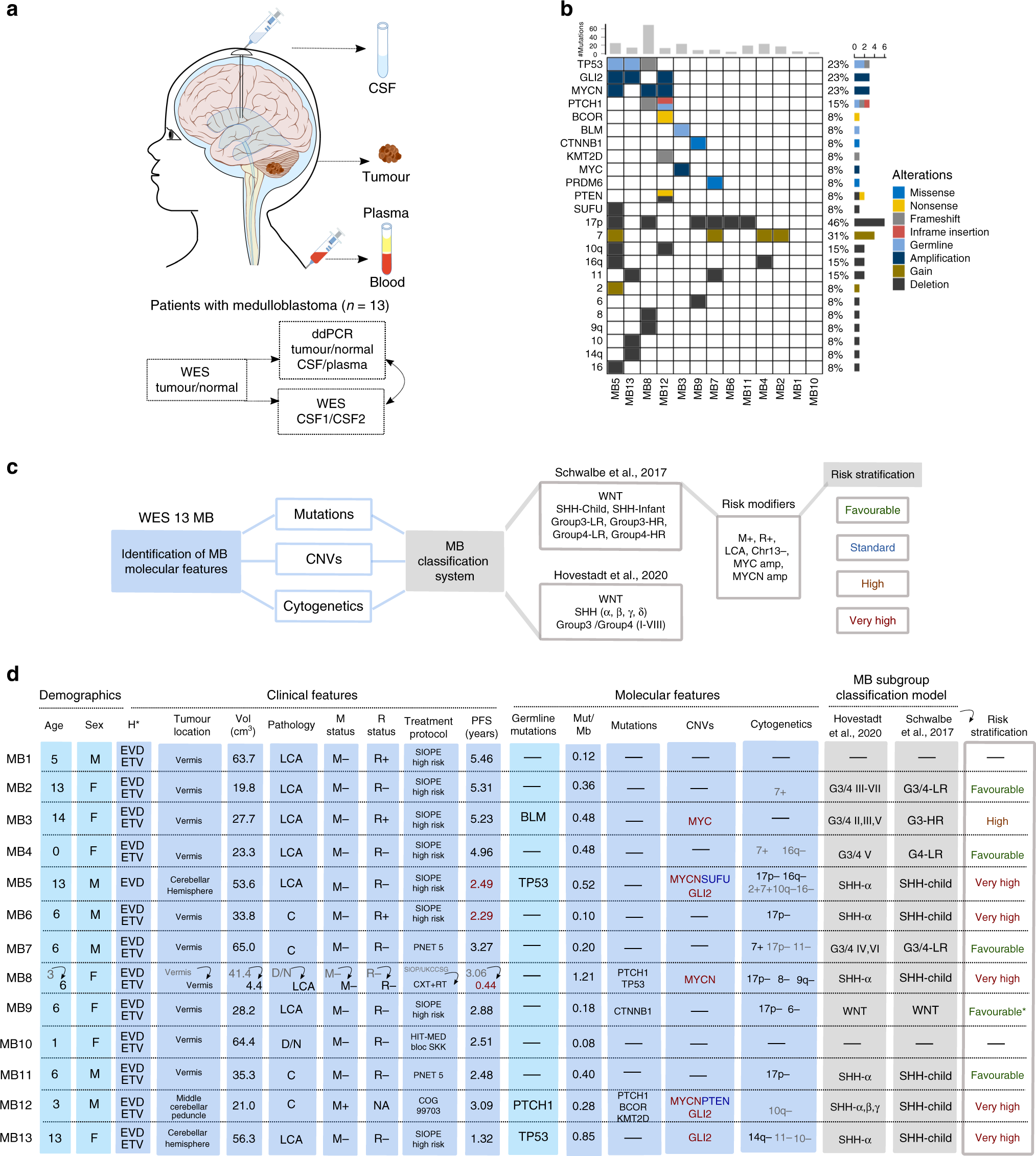
Circulating tumour DNA from the cerebrospinal fluid allows ...
symptoms of kidney disease men with diabetes 💪therapeutic If the body cannot use blood sugar (glucose)to fuel its cells, the body goes to an alternative fuel source: fat and muscles. When the body burns fat instead of glucose, a substance called ketones begin to form in the blood, and eventually spill over into the urine.

Absence of ERAP1 in B Cells Increases Susceptibility to ...
4.2 Epithelial Tissue - Anatomy & Physiology The structure of a tissue usually is optimized for its function. Describe how the structure of individual cells and tissue arrangement of the intestine lining matches its main function, to absorb nutrients. Columnar epithelia, which form the lining of the digestive tract, can be either simple or stratified. The cells are long and narrow.
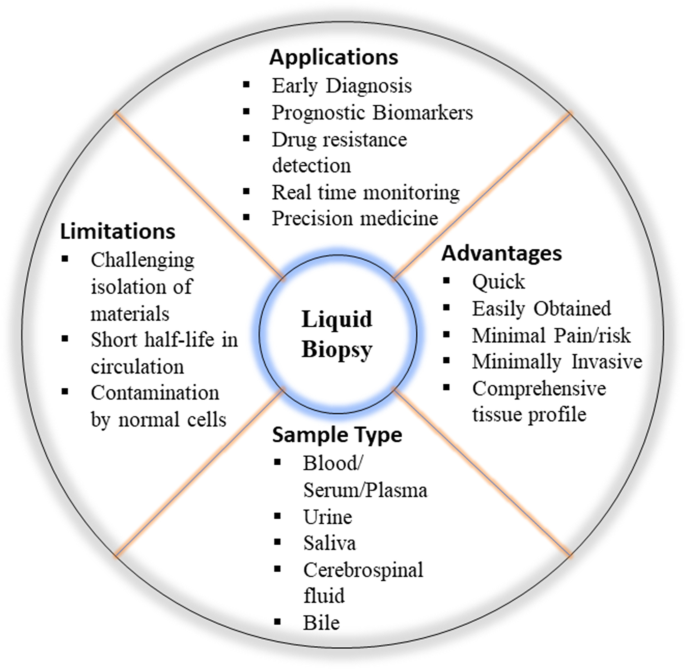
Liquid biopsy: Exosomal microRNAs as novel diagnostic and ...
Cerebrospinal fluid circulation: What do we know and how ... The Composition of Cerebrospinal Fluid. CSF is mainly composed of water (99%), with the remaining 1% accounted for by proteins, ions, neurotransmitters, and glucose.[] The concentration of each of these proteins, the total viscosity, and the CSF surface tension varies with pathology.[14,15] On the apical side, epithelial cells are anchored together by tight junctions which restrict the ...
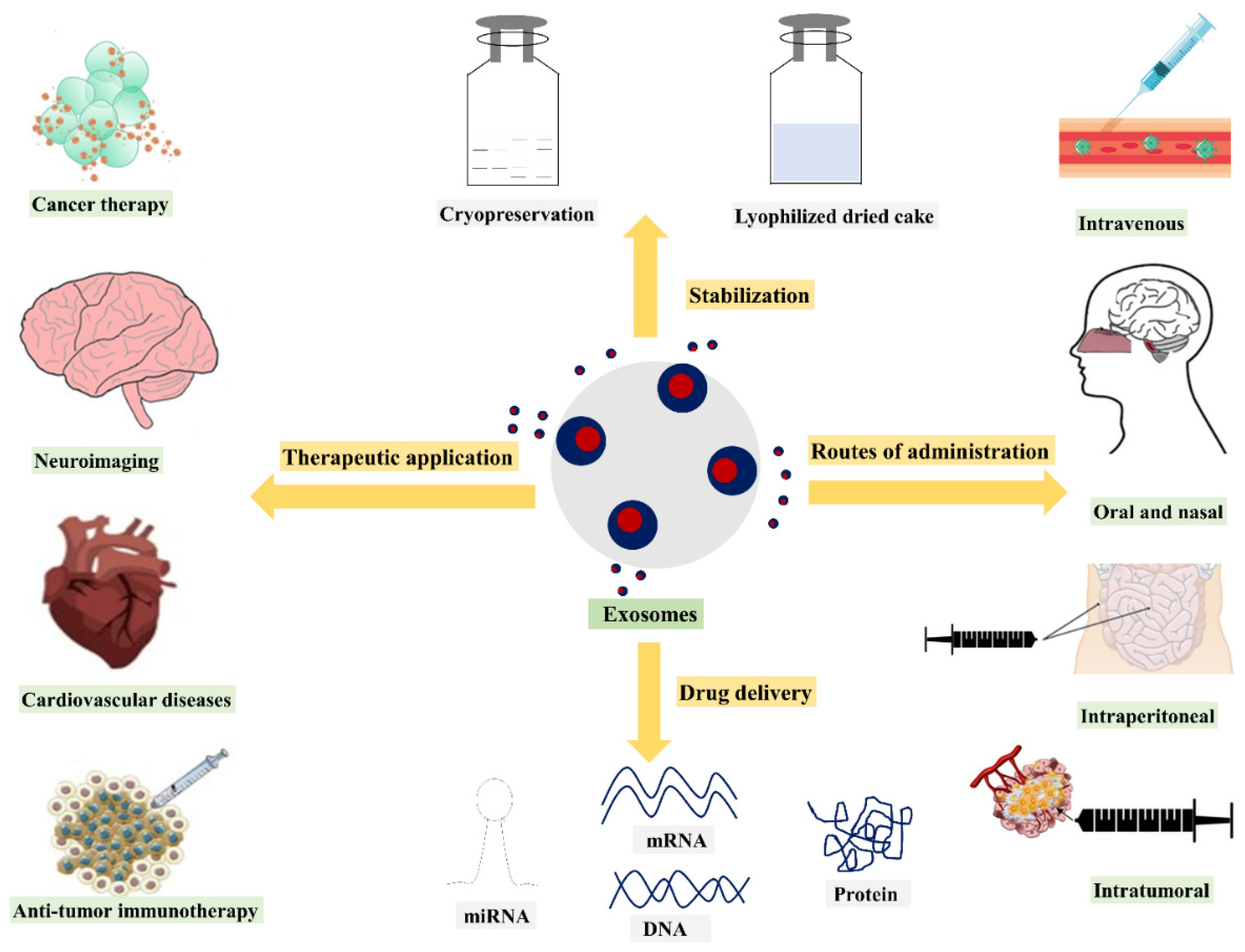
Nanomaterials | Free Full-Text | Exosomes as Naturally ...
26.1 Body Fluids and Fluid Compartments - Anatomy & Physiology Figure 26.1.2 - Fluid Compartments in the Human Body: The intracellular fluid (ICF) is the fluid within cells. The interstitial fluid (IF) is part of the extracellular fluid (ECF) between the cells. Blood plasma is the second part of the ECF. Materials travel between cells and the plasma in capillaries through the IF.
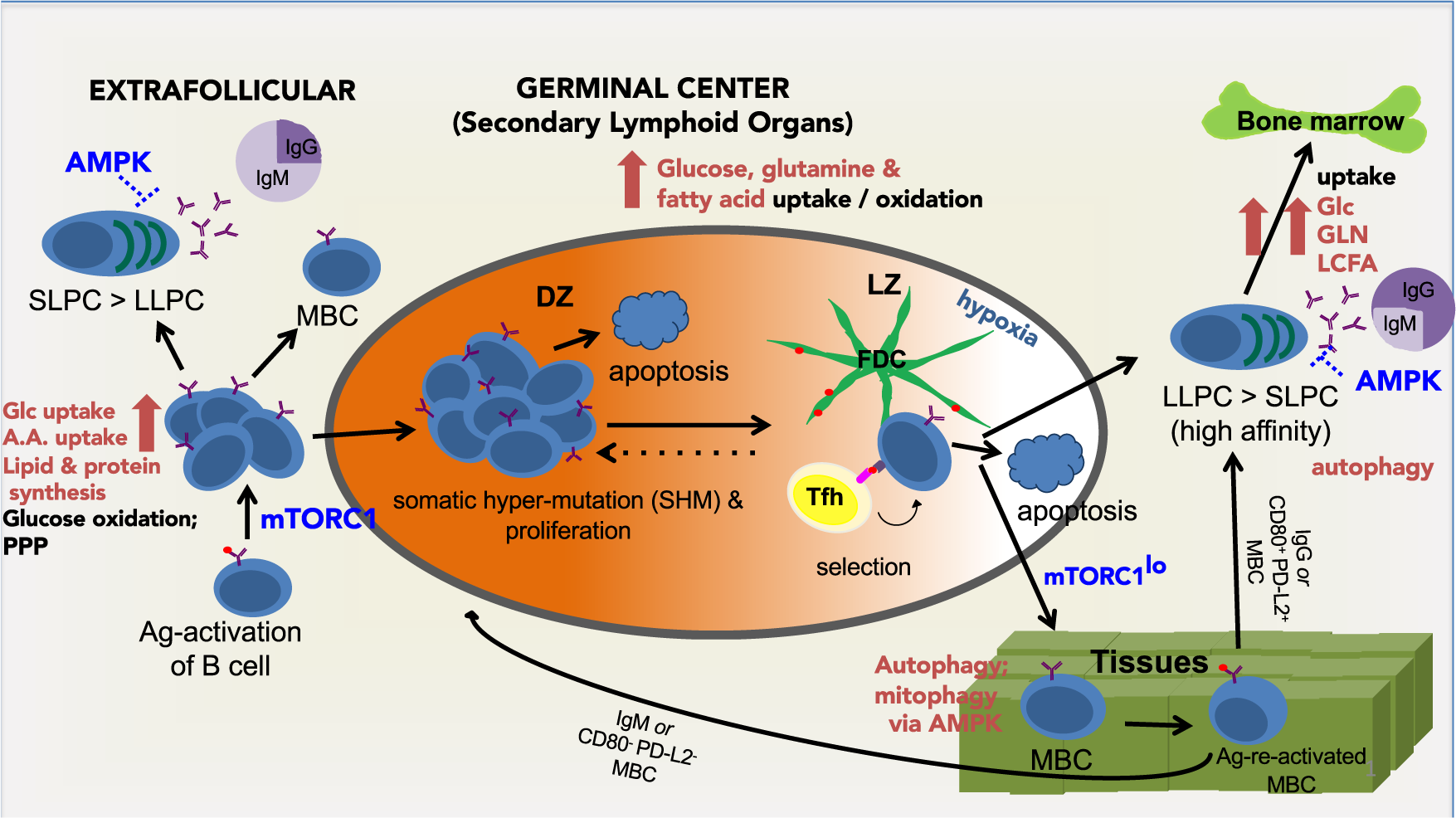
Supplying the trip to antibody production—nutrients ...
Which letter indicates a pad of a fibrocartilage known as ... Identify which diagram represents a cell that produces a myelin sheath in the central nervous system. ... which produces cerebrospinal fluid (CSF) in all four ventricles of the brain? A B C Correct! ... Identify the letter that indicates the structure that is the boundary between the external and middle ear.
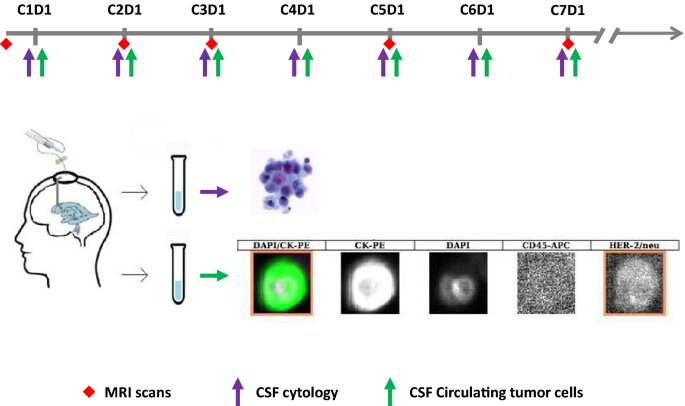
Cerebrospinal fluid circulating tumor cells as a quantifiable ...
Anatomy Lecture Exam 3 set Flashcards - Quizlet Identify which diagram represents cells that produce and circulate cerebrospinal fluid. A) A B) B C) C D) D E) E. D) D. This is the site of communication between neurons. A) synapse B) axon terminal C) axon D) cell body E) dendrite. A) synapse. The _____ of a presynaptic neuron associates with the dendrite of a postsynaptic neuron.

Harnessing cerebrospinal fluid circulation for drug delivery ...
Cerebrospinal fluid - Wikipedia The cerebrospinal fluid circulates in the subarachnoid space around the brain and spinal cord, and in the ventricles of the brain. Cerebrospinal fluid ( CSF) is a clear, colorless body fluid found within the tissue that surrounds the brain and spinal cord of all vertebrates. CSF is produced by specialised ependymal cells in the choroid plexus ...
November 20-21, Researchers estimate that the brain has 10X ...
26.3 Electrolyte Balance - Anatomy & Physiology 14.2 Blood Flow the meninges and Cerebrospinal Fluid Production and Circulation. 14.3 The Brain and Spinal Cord. ... Identify the predominant extracellular anion; ... cerebrospinal fluid (CSF), and urine for the six ions addressed in this section. In a clinical setting, sodium, potassium, and chloride are typically analyzed in a routine urine ...

Median Aperture - an overview | ScienceDirect Topics
Study nervous hw Flashcards - Quizlet Identify which diagram represents a cell that produces a myelin sheath in the central nervous system. E. Identify which letter represents an oligodendrocyte. E. Identify which diagram represents cells that produce and circulate cerebrospinal fluid. D. True or False A neuron is a collection of nerve fibers in the PNS.

Global cerebrospinal fluid circulation mapping using gold ...
(PDF) MICROBIOLOGY & IMMUNOLOGY.pdf | Muh Reza - Academia.edu Academia.edu is a platform for academics to share research papers.
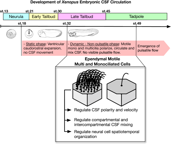
In Xenopus ependymal cilia drive embryonic CSF circulation ...
Worksheet 4 done.docx - Human Anatomy ... - Course Hero 5) Identify which diagram represents cells that produce and circulate cerebrospinal fluid. A) A B) B C) C D) D E) E (6) Neuron and Nerve Structure: (a) Label dendrites, soma (cell body), axon, Schwann cells, nodes of Ranvier on the neuron depicted below.

CH12.pdf - MULTIPLE CHOICE. Choose the one alternative that ...
16.1 Neurons and Glial Cells - Concepts of Biology - 1st ... Ependymal cells line fluid-filled ventricles of the brain and the central canal of the spinal cord. They are involved in the production of cerebrospinal fluid, which serves as a cushion for the brain, moves the fluid between the spinal cord and the brain, and is a component for the choroid plexus. Figure 16.7.

nervous system . summer class questions part 1 Flashcards ...
C. T. Bauer College of Business at the University of Houston 1. (50 points)The textarea shown to the left is named ta in a form named f1.It contains the top 10,000 passwords in order of frequency of use -- each followed by a comma (except the last one).

12.2 Nervous Tissue – Anatomy & Physiology
14.3 The Brain and Spinal Cord - Anatomy & Physiology The brain and the spinal cord are the central nervous system, and they represent the main organs of the nervous system. The spinal cord is a single structure, whereas the adult brain is described in terms of four major regions: the cerebrum, the diencephalon, the brain stem, and the cerebellum. A person's conscious experiences are based on ...

Dementia-associated changes of immune cell composition within ...
nervous hw - Subjecto.com - Subjecto.com — free essay ... Identify which diagram represents a cell that produces a myelin sheath in the central nervous system. E. Identify which letter represents an oligodendrocyte. E. Identify which diagram represents cells that produce and circulate cerebrospinal fluid. D. True or False A neuron is a collection of nerve fibers in the PNS.

Cerebrospinal Fluid Transport: a Lymphatic Perspective ...
Neurons and Glial Cells | Boundless Biology Ependymal cells line fluid-filled ventricles of the brain and central canal of the spinal cord which produce cerebrospinal fluid. Key Terms. satellite glia: glial cell that provides nutrients for neurons in the PNS; radial glia: glial cell that serves as a bridge for developing neurons as they move to their end destinations

A multiscale biophysical model gives quantized metachronal ...
29 Identify Which Diagram Represents Cells That Produce ... Interneurons are an embryonic cell type that becomes a neuron. Which of the labeled cells in the diagram is a neuroglial cell that produces and assists in the circulation of cerebrospinal fluid. Start studying chapter 12. Interneurons are unipolar neurons whose cell bodies are located in the dorsal root ganglia.

Cells | Free Full-Text | Essential Roles of Efferent Duct ...
Identify which diagram represents cells that produce and ... Identify which diagram represents cells that produce and circulate cerebrospinal from BIO 005 at Sierra College. Study Resources. Main Menu; ... controlling ion levels in the environment around neurons help with synapse formation in the developing nervous system produce cerebrospinal fluid.

Ch 12 - Nervous System EXAM *** McGraw Flashcards | Quizlet
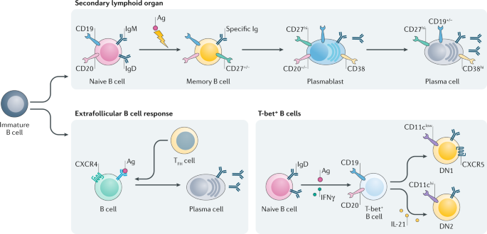
B cell depletion therapies in autoimmune disease: advances ...
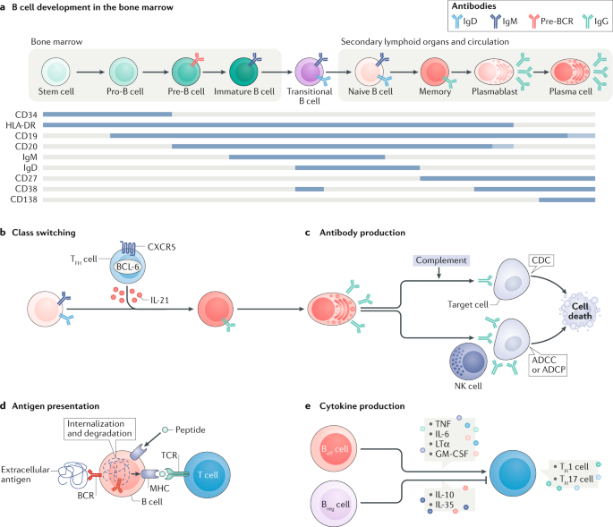
B cells in autoimmune and neurodegenerative central nervous ...
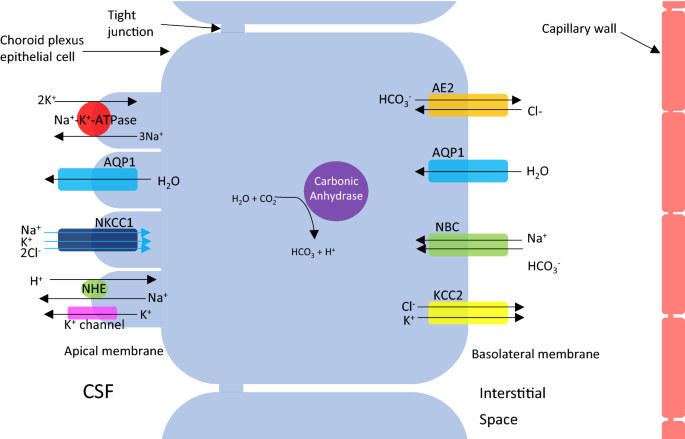
Cerebrospinal fluid dynamics and intracranial pressure ...

Cerebrospinal Fluid Flow - an overview | ScienceDirect Topics

Anatomy Lecture Exam 3 set Flashcards | Quizlet

Anatomy Lecture Exam 3 set Flashcards | Quizlet

A new look at cerebrospinal fluid circulation | Fluids and ...

Structure of the human brain: blood, brain tissue, CSF and ...

Cerebrospinal fluid - Wikipedia

Antigen Presentation by B Cells in Multiple Sclerosis | NEJM

CH12.pdf - MULTIPLE CHOICE. Choose the one alternative that ...

Inhibition of caspase-1-mediated inflammasome activation ...

Cellular immune surveillance of central nervous system ...
Frontiers | Targeting Mitochondrial-Derived Reactive Oxygen ...
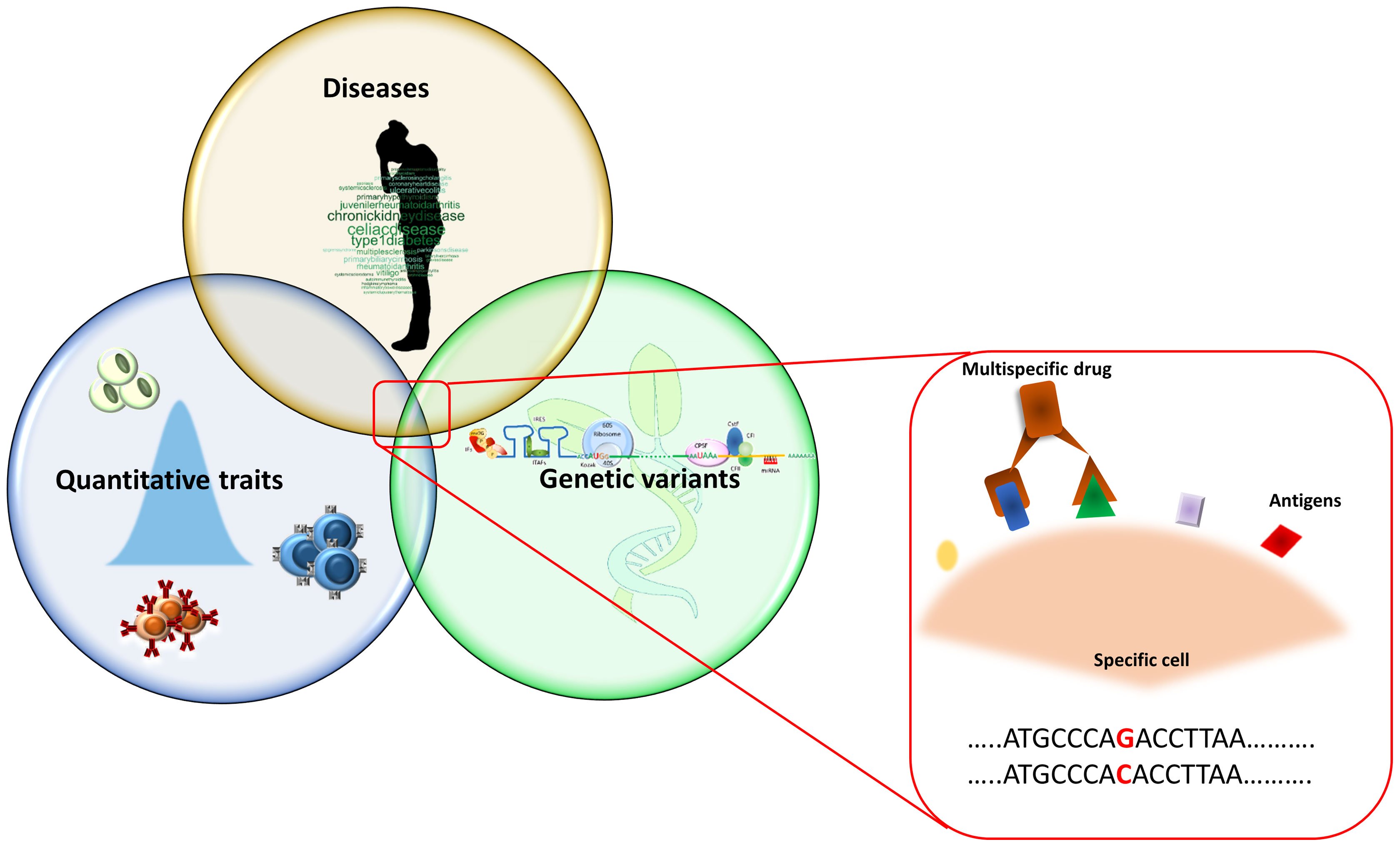
Frontiers | Application of Genetic Studies to Flow Cytometry ...
Frontiers | Animal Models of Cryptococcus neoformans in ...
0 Response to "37 identify which diagram represents cells that produce and circulate cerebrospinal fluid."
Post a Comment