36 human cheek cell diagram
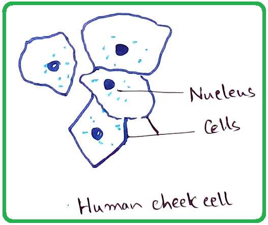
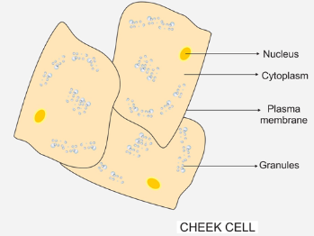
Unit: 4HB0 Paper: 01 - Edexcel 10.1.2019 · *P55799RA0328* 3 Turn over DO NOT WRITE IN THIS AREA DO NOT WRITE IN THIS AREA DO NOT WRITE IN THIS AREA (d) The diagram shows a human cheek cell. Lab 3: Cells: Structure and Function Animal Cells: Human Cheek Cells Prepare a wet mount of your cheek cells using the same technique you used last week. Observe the stained cells under high power. Find the nucleus, cytoplasm, and location of the cell membrane. Can you see any other organelles? If so, what might they be? Draw a well-labeled diagram of the cheek cells on the paper ...
Submandibular Lymph Nodes Anatomy, Diagram & Function ... 20.1.2018 · The submandibular lymph nodes sit between the submandibular salivary glands, which are underneath the tongue, and the mandible, or lower jawbone. Occasionally one or more of the lymph nodes may be ...
Human cheek cell diagram
Cell Nucleus - function, structure, and under a microscope ... [In this figure] The cell nucleus diagram and its structure. The nucleus consists of the nuclear envelope like double-layer membranes with pores ( nuclear pores), DNA, nucleolus (a site for ribosome synthesis, plural: nucleoli ), nucleoplasm (like cytoplasm of a cell), and the nuclear matrix, a supportive structure like the cytoskeleton supports cells. Human Cell Diagram, Parts, Pictures, Structure and Functions Feb 06, 2017 · Human Cell Diagram, Parts, Pictures, Structure and Functions The cell is the basic functional in a human meaning that it is a self-contained and fully operational living entity. Humans are multicellular organisms with various different types of cells that work together to sustain life. Human Cheek Epithelial Cells - Florida State University Nov 13, 2015 · Human Cheek Epithelial Cells. The tissue that lines the inside of the mouth is known as the basal mucosa and is composed of squamous epithelial cells. These structures, commonly thought of as cheek cells, divide approximately every 24 hours and are constantly shed from the body.
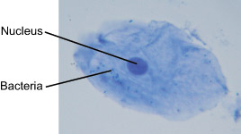
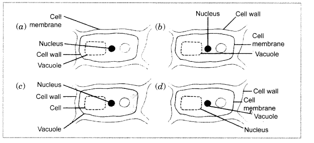
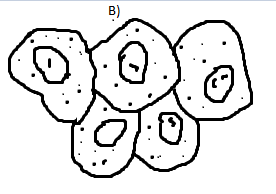
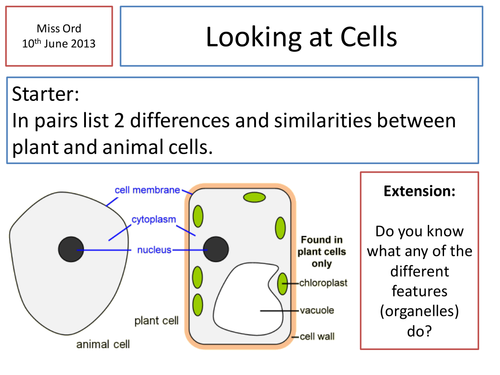
Human cheek cell diagram. Human Cheek Cell Diagram Labeled - Diagram Sketch Sep 18, 2021 · Human Cheek Cell Diagram Labeled. angelo on September 18, 2021. Label The Plant Cell Worksheets Sb11867 Sparklebox Cells Worksheet Plant Cell Plant Cells Worksheet. Art Animal Cells Do Not Have Cell Walls They Can Change Size And Shape More Easily Than Plant Cells Animal Cell Plant And Animal Cells Animal Cell Project. Which of the following is the diagram of human cheek cell? A cell membrane that is semi-permeable surrounds the cytoplasm. The vacuole in an animal cell is smaller in size, or absent. The nucleus is present at the centre of the cytoplasm. The absence of a cell wall and a prominent vacuole are indicators that help identify animal cells, such as cells seen in the human cheek. Comparing sizes - Cell structure - AQA - GCSE Combined ... The diagram shows the size of ... What is the width of a cheek cell compared with a Salmonella ... Meaning there is one order of magnitude between the height of a human being (2m) and the ... Human Cheek - Experiments on Microscopes 4 Schools Human cheek cells experiment from Microscopes for Schools. Observing cells from a human cheek and bacteria under a compound microscope.
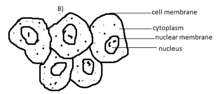
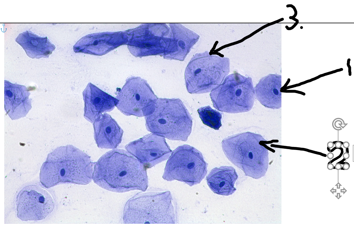
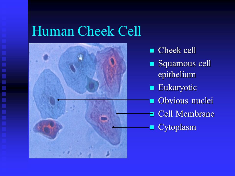
Human cheek cell diagram labeled Human cheek cell diagram labeled. Extra Questions "Why was the wicking procedure used?" Wicking draws stain through a tissue on a slide instead of dropping ... Human Cheek Cells Under the Microscope | Haematoxylin | Cell ... Human cheek cells are observed under microscope-1. Cells are polygonal or flat in shape and structure – 2. They have irregular cellular thin boundaries which contains jelly like cytoplasm and the cytoplasm are granular. 3. This cell do not have plastids, vacuoles or cell wall. 4. They are generally made up of squamous epithelium cells. Human Cheek Epithelial Cells - Florida State University Nov 13, 2015 · Human Cheek Epithelial Cells. The tissue that lines the inside of the mouth is known as the basal mucosa and is composed of squamous epithelial cells. These structures, commonly thought of as cheek cells, divide approximately every 24 hours and are constantly shed from the body. Human Cell Diagram, Parts, Pictures, Structure and Functions Feb 06, 2017 · Human Cell Diagram, Parts, Pictures, Structure and Functions The cell is the basic functional in a human meaning that it is a self-contained and fully operational living entity. Humans are multicellular organisms with various different types of cells that work together to sustain life.
Cell Nucleus - function, structure, and under a microscope ... [In this figure] The cell nucleus diagram and its structure. The nucleus consists of the nuclear envelope like double-layer membranes with pores ( nuclear pores), DNA, nucleolus (a site for ribosome synthesis, plural: nucleoli ), nucleoplasm (like cytoplasm of a cell), and the nuclear matrix, a supportive structure like the cytoskeleton supports cells.
Human Cheek Cells Under the Microscope | Haematoxylin | Cell ...
Human Cheek - Experiments on Microscopes 4 Schools

The Human Cheek Cell by Jacob Fritz
CBSE Class 9 Science Practical Skills – Slide of Onion Peel ...
The Human Cheek Cell
The following diagram shows cells of onion peel label class ...

Lab: Observing Cells – Michael Eng – Michael's Blog
Solved a). Indicate by placing a tick beside the description ...

Effect of mouthwashes on the morphology of human cheek cells ...
Human Cells and Microscopy

NCERT Class 9 Science Lab Manual - Slide of Onion Peel and ...
How to draw #human cheek cell || Most easy way || Step by ...
The following diagram shows cells of onion peel label class ...

Which of the following is the diagram of human cheek cell?

Cell Division. - ppt download
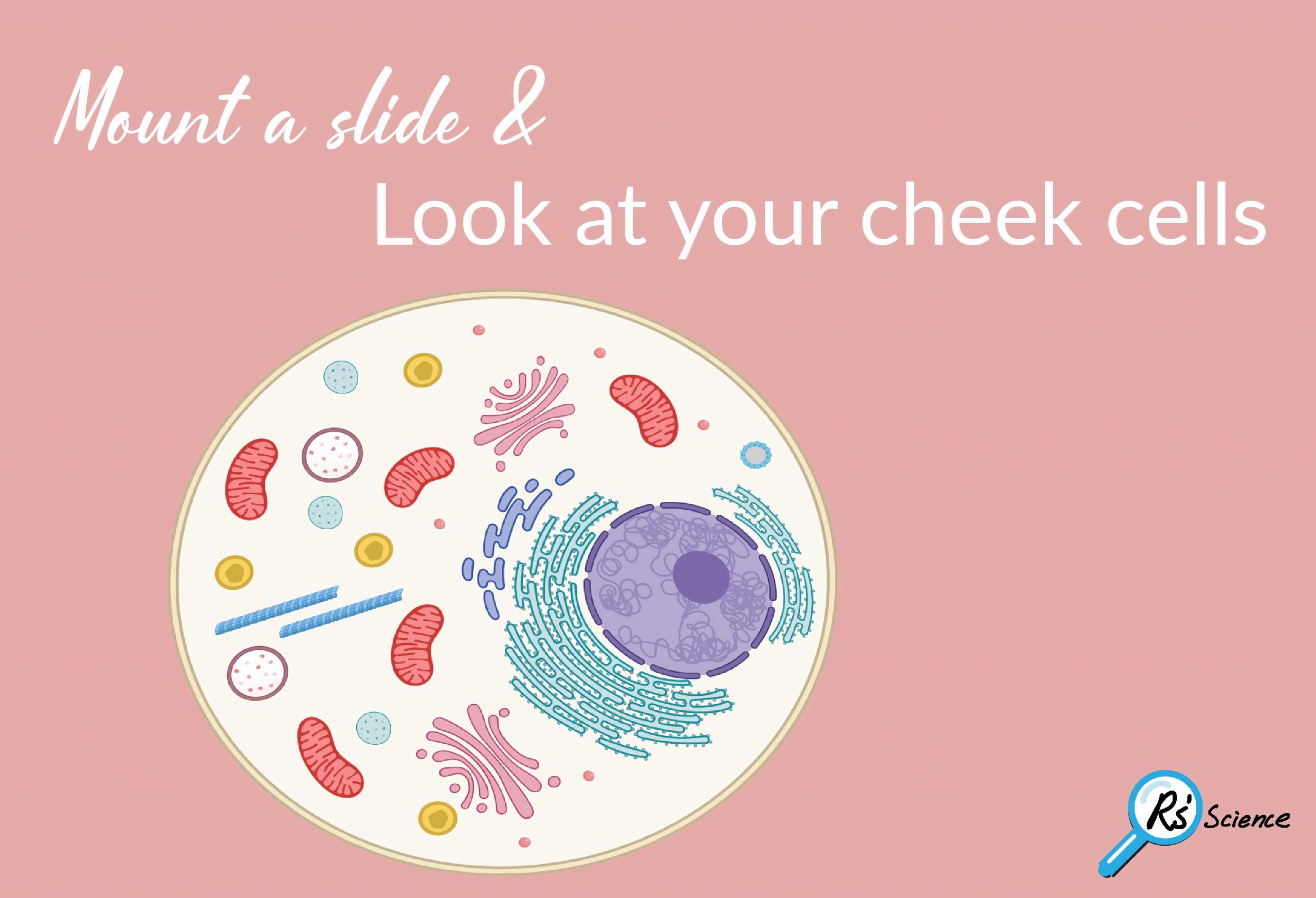
Lesson 2: Mount a Slide & “Look at Your Cheek Cells“ - Rs ...

Draw the diagram of cheek cells and label the parts. - Brainly.in

Cheek cell image using brightfield and darkfield microscopy ...

Cheek cell image using brightfield and darkfield microscopy ...
What are the benefits of apoptosis to a eukaryotic cell? - Quora
POST LAB: Bacterium, Plant & Animal CELLS - ppt download

HUMAN CHEEK CELL ( Class : 8 Lesson No : 8 )

Draw a diagram of cheek cells and it's two identifying points ...

Images of human epithelial cheek cells. (a) Image recorded by ...

Barr body in Cheek cells – Microcosmos

Day 18 What do a paramecium and a human cheek cell have in ...
Cheek cells practical | Teaching Resources
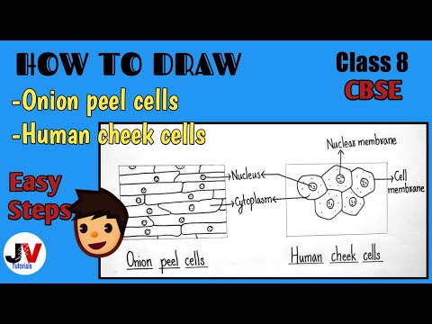
How to draw human cheek cells| How to draw onion peel cells|NCERT

Cell organelle present in both prokaryotic and eukaryotic ...
Figure 1 shows a human cheek cell viewed under a light ...
Which of the following represents the human cheek class 11 ...

how to draw human cheek cell diagram | how to draw cheek cell ...

Human Cheek Cells and Plant Cells

abril | 2015 | Discovering Life in class
investigating human cheek cell ss

Cheek Cell Investigation
0 Response to "36 human cheek cell diagram"
Post a Comment