40 veins of the head and neck diagram
May 14, 2018 Anatomy, Head and Neck external carotid artery, external jugular vein, internal jugular vien, MCQs on head and neck, Muscles of mastication, nerve supply of tongue, parotid gland, scap dangerous layer POONAM KHARB JANGHU Jul 29, 2020 · The cardiovascular system consists of the heart, blood vessels, and the approximately 5 liters of blood that the blood vessels transport. Responsible for transporting oxygen, nutrients, hormones, and cellular waste products throughout the body, the cardiovascular system is powered by the body’s hardest-working organ — the heart, which is …
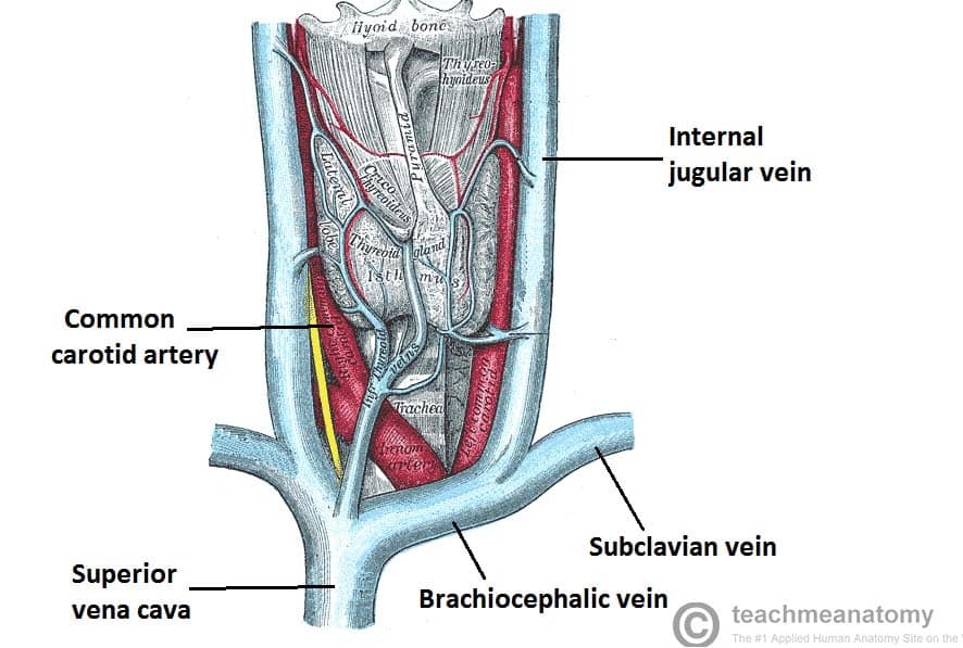
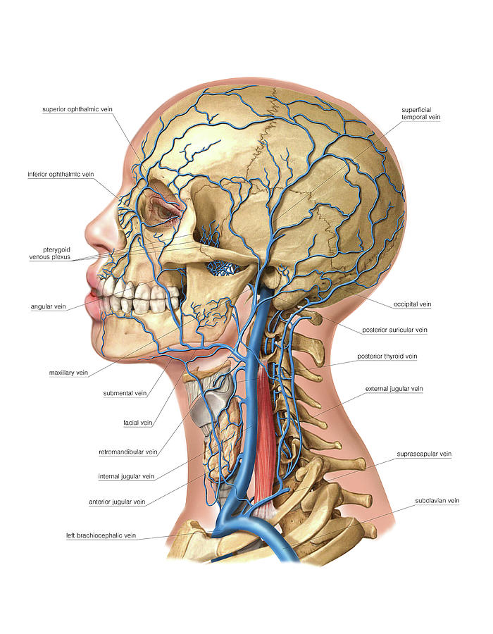
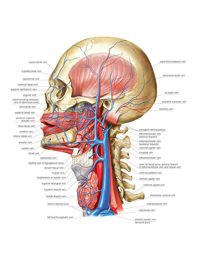
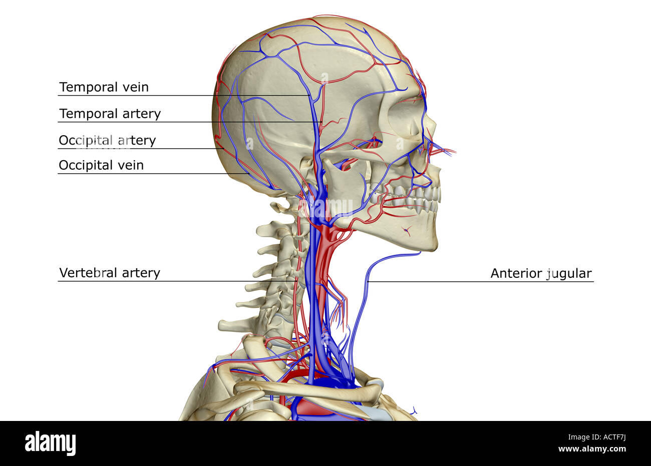
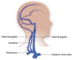
Dec 21, 2021 · Veins and arteries of the head (a diagram) Deoxygenated blood from the brain, head, and neck ultimately drain into one of the three jugular veins: external, internal, or anterior. Venous blood of the brain and meninges drains into the dural venous sinuses, which drain into the internal jugular vein.

Veins of the head and neck diagram
FIG.557– Veins of the head and neck. The veins of the head and neck may be subdivided into three groups: (1) The veins of the exterior of the head and face. (2) The veins of the neck. (3) The diploic veins, the veins of the brain, and the venous sinuses of the dura mater.: 1. The Veins of the Exterior of the Head and Face—The veins of the exterior of the head and face (Fig. 557) are: Carotid artery angiography, known as an angiogram: Contrast dye is injected into blood vessels, and X-rays are taken of the neck, revealing images of the carotid arteries. A narrowing, or stenosis ... Jun 11, 2021 · The salivary glands are exocrine glands that make, modify and secrete saliva into the oral cavity. They are divided into two main types: the major salivary glands, which include the parotid, submandibular and sublingual glands, and the minor salivary glands, which line the mucosa of the upper aerodigestive tract and the overwhelming entirety of the mouth [1].
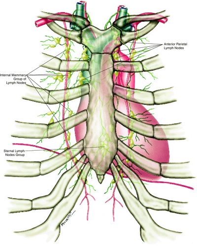
Veins of the head and neck diagram. Neck muscles are bodies of tissue that produce motion in the neck when stimulated. The muscles of the neck run from the base of the skull to the upper back and work together to bend the head and ... #anatomy #jugular #carotidhttps://www.instagram.com/anatomy.knowledge/Veins of the head and neck: Jugular veins and their tributaries.Facial vein, pterigoid ... 3b. 2. The Veins of the Neck - Human Anatomy. FIG.558– The veins of the neck, viewed from in front.(Spalteholz.) The veins of the neck (Fig. 558), which … 5) Regarding lymph drainage of head and neck, which is INCORRECT? a) superficial cervical nodes lie along the external jugular veins b) submandibular nodes are lateral to submental nodes c) the left jugular lymph trunk usually drains into the thoracic duct d) the jugulodigastric nodes lie on/near the tendon of omohyoid
The vorticose veins, referred to clinically as the vortex veins, drain the ocular choroid.The number of vortex veins is known to vary from 4 to 8 with about 65% of the normal population having 4 or 5. In most cases, there is at least one vortex vein in each quadrant. Insect morphology is the study and description of the physical form of insects.The terminology used to describe insects is similar to that used for other arthropods due to their shared evolutionary history. Three physical features separate insects from other arthropods: they have a body divided into three regions (called tagmata) (head, thorax, and abdomen), have three … Nov 03, 2021 · Overview. The veins of the brain are thin-walled, valveless and pierce the arachnoid mater and meningeal layer of dura mater (of meninges) to empty poorly oxygenated blood into the dural venous sinuses.The dural venous sinuses drain into the sigmoid sinus which becomes continuous with the internal jugular veins (IJVs). Feb 09, 2017 · Enumerate: Layers of scalp; Sensory nerves supplying scalp.; Arteries supplying scalp.; Arteries supplying face.; Components of lacrimal apparatus.; Structures pierces by p arotid duct.; Structures passing through the parotid gland.; Branches of f acial artery.; Cutaneous nerve of side of neck and their root values.; Tributaries of external jugular vein; Contents of posterior …
Head and neck (anterior view) The head and neck are two examples of the perfect anatomical marriage between form and function, mixed with a dash of complexity. The neck is resilient enough to sustain a five kilogram weight 24/7, yet sufficiently mobile to move it in several directions. Veins of the Head and Neck: The veins of head and neck are labeled in blue, with an arrow pointing to the auricular vein. The superficial external jugular vein is formed from the retromandibular vein and the posterior auricular vein at a point adjacent to the mandible. Jul 18, 2016 · The external jugular vein (v. jugularis externa) (Figs. 12-1 though 12-6, 12-8, and 12-10) is the main channel for return of venous blood from the head.It begins by the union of the linguofacial and maxillary veins, caudal to the mandibular salivary gland or at a transverse plane through the cricoid cartilage and the axis. Official Ninja Nerd Website: https://ninjanerd.org/Ninja Nerds!In this lecture Professor Zach Murphy will be presenting on the veins of the head and neck thr...
Vessel Anatomy: Veins of the Head and Neck. Diagram | Quizlet
Facial Anatomy. Head Anatomy. Muscle Anatomy. Anatomy Drawing. Facial Muscles. Blood Vessels Anatomy. Life Of Medicine’s Instagram photo: “Wow! 😱Look at the amazing detail of the anatomy of the human face! Showing the muscles, blood vessels, and nerves of the face! 🙌🏼 . .…”. 4,693 Likes, 32 Comments - Life Of Medicine (@life_of ...
:max_bytes(150000):strip_icc()/heart-and-circulatory-system-with-blood-vessels--97537745-a3bc2b2a6ca94390bfdf2696ad9bbddd.jpg)
Pulmonary Vein: Anatomy, Function, and Significance
Jun 11, 2021 · The salivary glands are exocrine glands that make, modify and secrete saliva into the oral cavity. They are divided into two main types: the major salivary glands, which include the parotid, submandibular and sublingual glands, and the minor salivary glands, which line the mucosa of the upper aerodigestive tract and the overwhelming entirety of the mouth [1].

veins of the head and neck Diagram | Quizlet
Carotid artery angiography, known as an angiogram: Contrast dye is injected into blood vessels, and X-rays are taken of the neck, revealing images of the carotid arteries. A narrowing, or stenosis ...
Venous Drainage of the Head and Neck - Dural Sinuses ...
FIG.557– Veins of the head and neck. The veins of the head and neck may be subdivided into three groups: (1) The veins of the exterior of the head and face. (2) The veins of the neck. (3) The diploic veins, the veins of the brain, and the venous sinuses of the dura mater.: 1. The Veins of the Exterior of the Head and Face—The veins of the exterior of the head and face (Fig. 557) are:

79 Neck Vein High Res Illustrations - Getty Images
Chap 18 – Blood Vessels Continued Learning Objectives ...

Lateral view of the dissected right side of the head and neck ...

CLASS BLOG: July 2012 | Arteries and veins, Arteries anatomy ...

Examination of the Neck Veins | NEJM
Major arteries, veins and nerves of the body: Anatomy | Kenhub

Veins from the Head Neck and Brain - Blood Vessels - GUWS Medical
Anatomy diagram showing crucial veins in human head and neck ...
The blood supply of the head and neck - Stock Photo ...

Circulation and the Central Nervous System | Anatomy and ...
Nerves and arteries of head and neck: Anatomy, branches | Kenhub
Internal jugular vein: Origin, course, drainage, JVP | Kenhub

Veins of the Head and Neck Diagram | Quizlet
Venous System Of The Head And Neck by Asklepios Medical Atlas

VEINS of the head and neck Diagram | Quizlet
/heart-and-circulatory-system-with-blood-vessels--97537745-a3bc2b2a6ca94390bfdf2696ad9bbddd.jpg)
Pulmonary Vein: Anatomy, Function, and Significance
Venous System Of The Head And Neck by Asklepios Medical Atlas

Venous system of the head and neck - Stock Image - C021/2137 ...

Circulatory Pathways | Anatomy and Physiology II
Veins of the Head and Neck | Radiology Key

Head and neck region - Knowledge @ AMBOSS
Veins of the head and neck - Labelled diagram

Circulatory Pathways | Anatomy and Physiology II
Temporal Artery High Resolution Stock Photography and Images ...
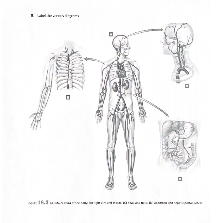
Solved B. Label the venous diagrams FIGURE 18.2 (A) Major ...

Major Veins of the head and neck Quiz
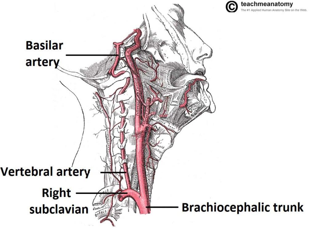
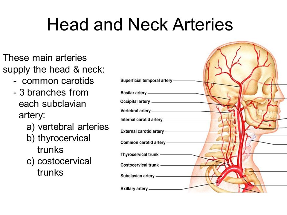
Major Arteries of the Head and Neck - Carotid - TeachMeAnatomy
/GettyImages-530309436-cf8e158016cf4dc0a81e12ecb221d1ee.jpg)
Internal Jugular Vein: Anatomy, Function, and Significance

me 5.12 by writing your answers in 2 Veins superior to the ...
Vasculature of the Head | Texas Heart Institute

8: Systemic Anatomy of the Head and Neck | Pocket Dentistry

Veins of the Head and Neck Diagram | Quizlet

Veins of the Head and Neck (right aspect) Quiz

Carotid artery: Anatomy, function, disease, and more
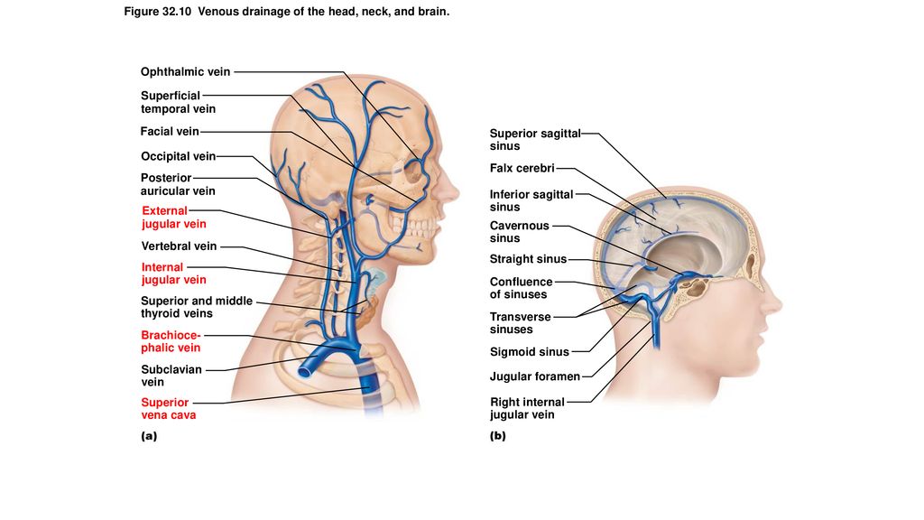
Figure 32.3a Arteries of the head, neck, and brain. - ppt ...
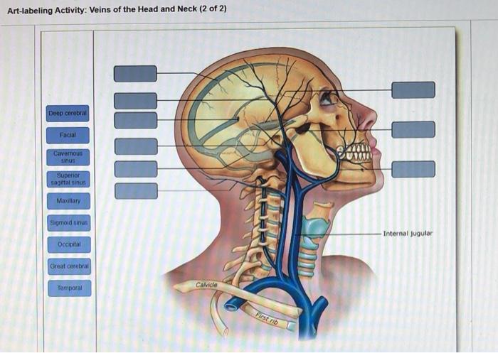
Solved Art-labeling Activity: Veins of the Head and Neck (2 ...
0 Response to "40 veins of the head and neck diagram"
Post a Comment