35 muscles of respiration diagram
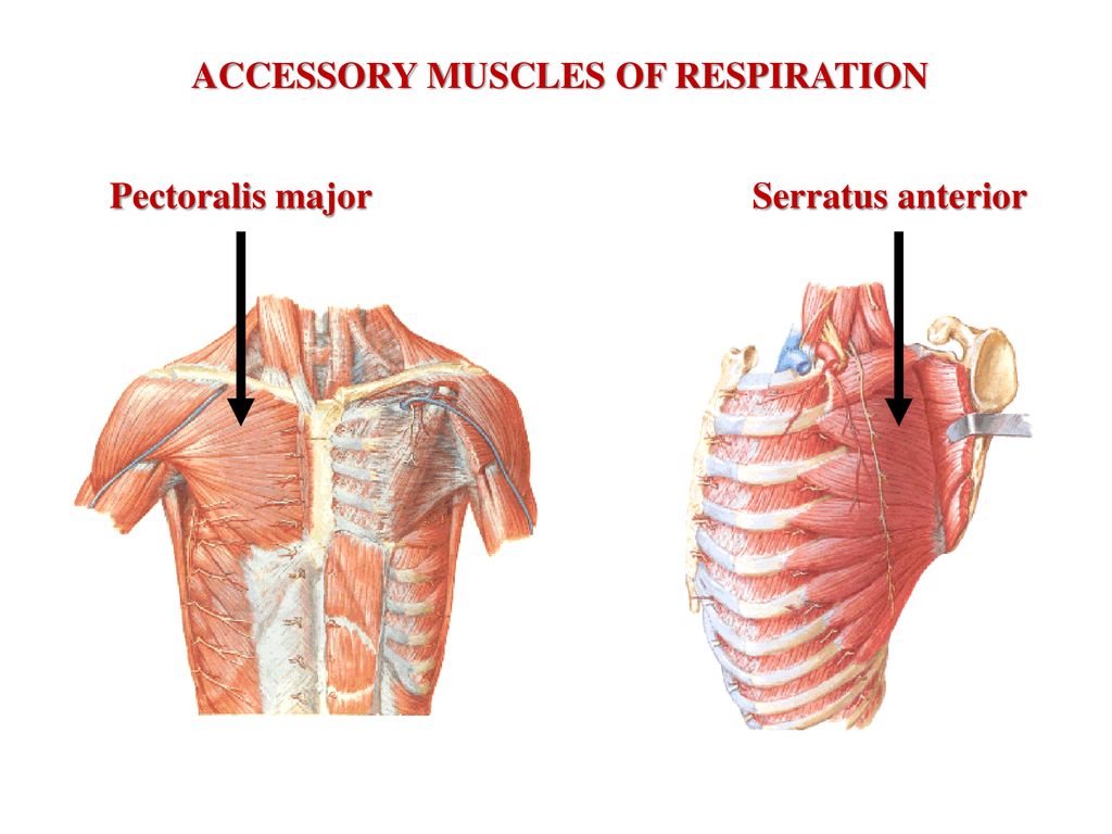
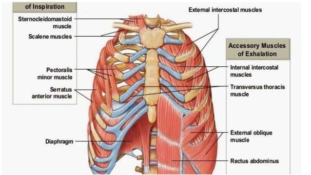
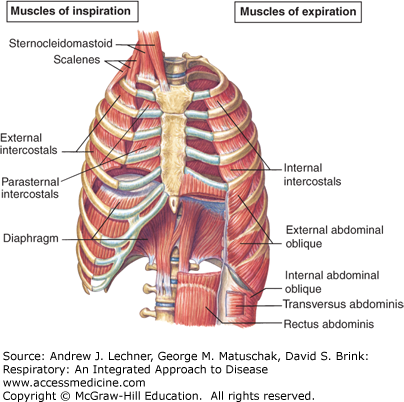
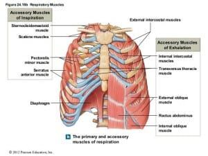
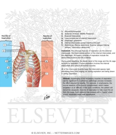
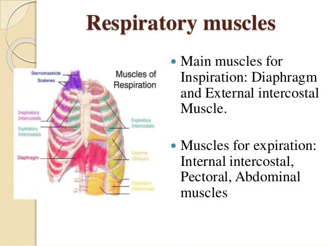
Muscles of Respiration Pleurae & Pleural cavitiesPleurae & Pleural cavities Respiratory Movements of the Chest -- Inspiration Inspiration It requires expansion of the t horax and increase of the: - Anteroposterior diameter of the thoracic chest During forced inspiration and expiration many other muscles besides the diaphragm and intercostal muscles come into play to assist respiration. Most important of this group are the scalene and sternocleidomastoids of the anterior neck muscles which stabilise the first ribs and upper sternum during forced inspiration.
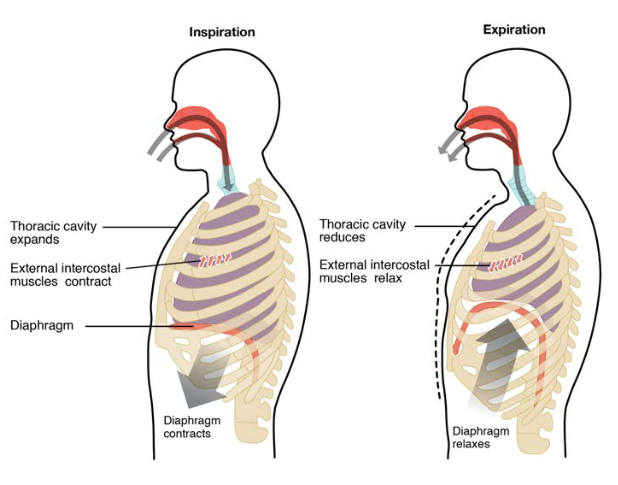
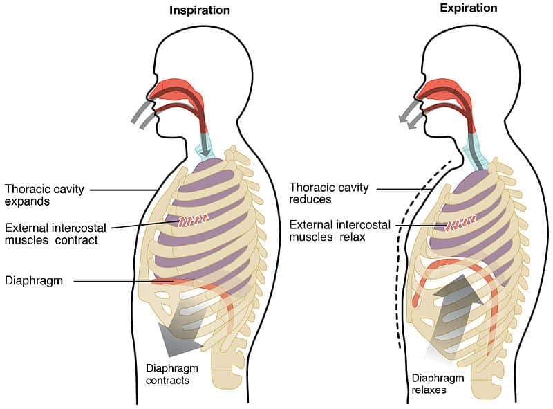
Muscles of Respiration During quiet breathing, the predominant muscle of respiration is the diaphragm. As it contracts, pleural pressure drops, which lowers the alveolar pressure, and draws air in down the pressure gradient from mouth to alveoli.
Muscles of respiration diagram
these muscles play a role in lifting up upper part of rib cage, have a small role in respiration scalenus muscle anterior, middle, and posterior muscles of the neck that elevate the first and second ribs accessory muscles of inspiration; support head scalenus muscles sternomastoid muscles these muscles will move when you have a shortage of breathe The muscles of respiration are also called the 'breathing pump muscles', they form a complex arrangement in the form of semi-rigid bellows around the lungs. All muscles that are attached to the human rib cage have the inherent potential to cause a breathing action. Muscles that helpful in expanding the thoracic cavity are called the inspiratory ... Bird respiratory system organs diagram. I have already shown you the important labelled diagram from the bird respiratory system. Here, I tried to show you the detailed anatomical facts of most organs from a bird (chicken). If you want to get a more detailed diagram and high-quality image, make sure you join with anatomy learner on social media.
Muscles of respiration diagram. The lungs are the main component of the human bodys respiratory system. Every healthy human body has two kidneys the left and the right. Learn about lung function problems location in the body and more. The interactive BodyMap diagram below shows the location of the heart in the body. Aside from the lungs there are also muscles and a vast. Start studying Respiratory--Anatomy, Muscles of respiration. Learn vocabulary, terms, and more with flashcards, games, and other study tools. 25 The diagram shows the bones and muscles of the upper arm. X Y ... D photosynthesis and respiration 36 The diagram shows the movement of carbon atoms in part of the carbon cycle. The directions of movement are not shown. carbon dioxide in the atmosphere fossil fuels bacteria See an arm muscle diagram to learn about arm muscle anatomy. Explore the parts of arm muscle and discover the purpose and function of each part. ... Go to Human Respiratory System Ch 17.
Diagram; Steps; Key Points; Respiration is of two types, aerobic respiration, and anaerobic respiration. Aerobic Respiration: It is the process of cellular respiration that takes place in the presence of oxygen gas to produce energy from food. This type of respiration is common in most of the plants and animals, birds, humans, and other mammals. Fig: Respiratory System-Elaborating the Internal Structure of Lung. Mechanism (Physiology) of Respiration. The entire Mechanism of Respiration involves the following steps:. Breathing (Pulmonary ventilation): The mechanism of breathing involves the inspiration and expiration of air with the movement of the diaphragm and intercostal muscles.During inhalation, external intercostal muscles contract. The respiratory tract in humans is made up of the following parts: External nostrils - For the intake of air.; Nasal chamber - which is lined with hair and mucus to filter the air from dust and dirt.; Pharynx - It is a passage behind the nasal chamber and serves as the common passageway for both air and food.; Larynx - Known as the soundbox as it houses the vocal chords, which are ... Start studying Quiz 2 - Internal & External Intercostal Muscles of Respiration. Learn vocabulary, terms, and more with flashcards, games, and other study tools.
Title: MUSCLES INVOLVED IN RESPIRATION Author: Dr. Zeenat Zaidi Last modified by: 3422 Created Date: 1/27/2010 8:25:16 AM Document presentation format - A free PowerPoint PPT presentation (displayed as a Flash slide show) on PowerShow.com - id: 780459-ZmRkM What muscles are used in the expiratory process? Inspiration or inhalation is the process of bringing air from outside the body into the lungs. It is carried out by creating a pressure gradient between the lungs and the atmosphere. When air enters the lungs, the diaphragm contracts toward the abdominal. cavity, thereby increasing the space in the thoracic cavity for accommodating the inhaled air. Jul 29, 2020 · The diaphragm is a dome-shaped sheet of muscle located below the lungs. It separates the chest from the abdomen. The diaphragm operates as the major muscle of respiration and aids breathing .
Yoga Muscles. Shiatsu. Lower Back Muscles. Muscle Anatomy. 10 of the Best Workouts for Weight Loss. If your aim is to lose weight, these 10 workouts are all excellent places to start. Find out how to exercise (and eat, and other things) to reach your goals. hebaguma8022. H.
Start studying Respiratory: muscles. Learn vocabulary, terms, and more with flashcards, games, and other study tools.
Respiratory System Diagram Muscles of the Respiratory System. The diaphragm is a dome-shaped muscle that curves upwards towards the lungs. When it contracts, it becomes flattened and therefore increases the volume of the thoracic cavity.
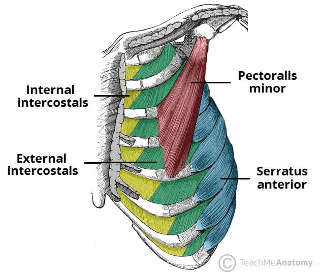
Inspiration is the phase of ventilation in which air enters the lungs. It is initiated by contraction of the inspiratory muscles: Diaphragm - flattens, extending the superior/inferior dimension of the thoracic cavity. External intercostal muscles - elevates the ribs and sternum, extending the anterior/posterior dimension of the thoracic cavity.
Oct 09, 2021 · Respiratory system (anatomy diagram) So far, you have seen how the thoracic cage is a frame that encloses the respiratory system and allows breathing to take place. Several muscles that span several regions of the body, such as the thoracic wall itself, neck, shoulder girdle and abdomen , act upon this structure.
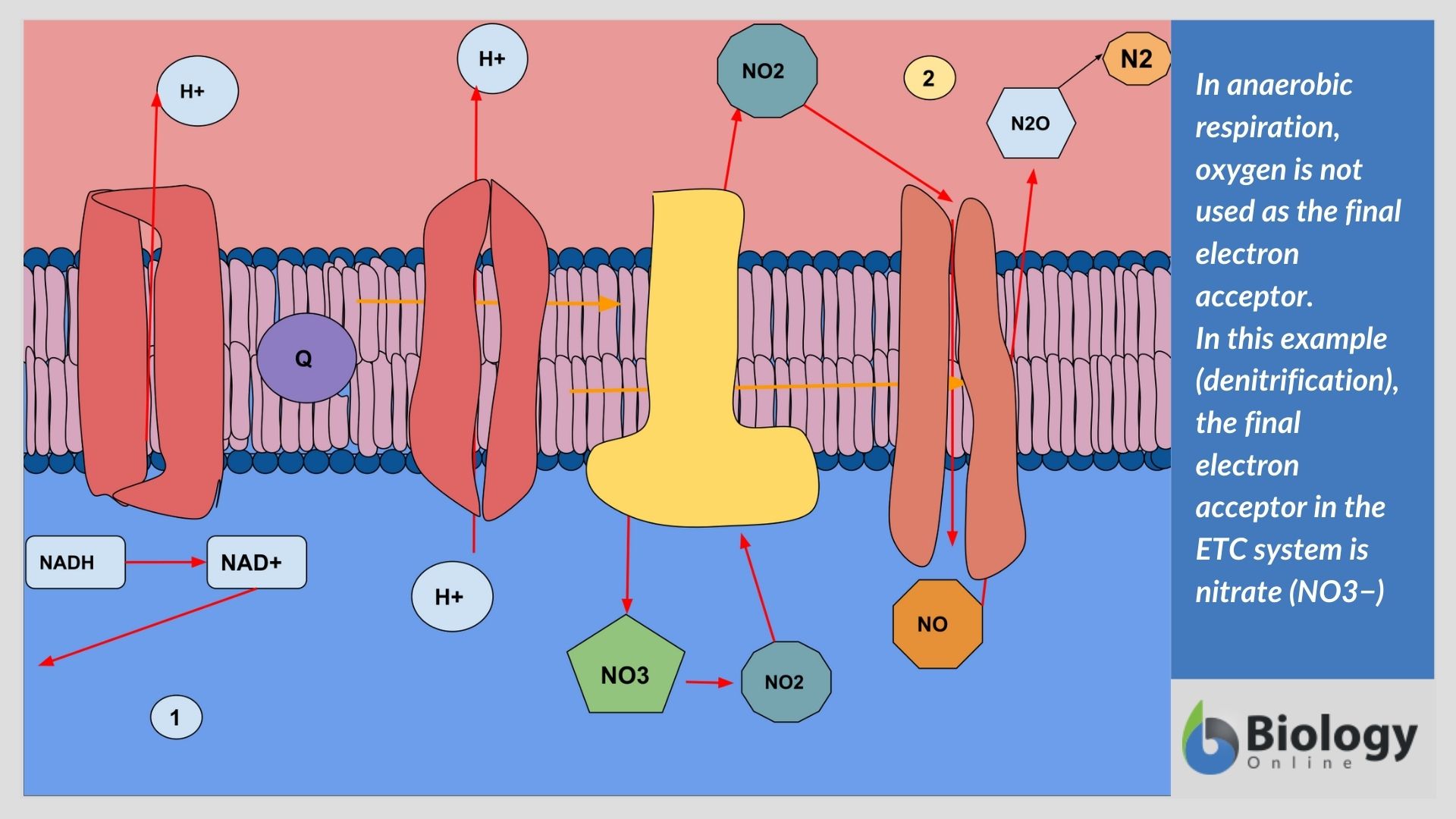
Anaerobic Respiration (With Diagram) Anaerobic respiration is an alternate mode of energy generation in which an exogenous electron acceptor other than O 2 is used in electron transport chain leading to a proton motive force. In contrast to aerobic respiration where O 2 is used as electron acceptor, the electron acceptors used in anaerobic ...
The diaphragm is the primary muscle used in respiration, which is the process of breathing. This dome-shaped muscle is located just below the lungs and heart. It contracts continually as you ...
Muscles get their energy from different sources depending on the situation that the muscle is working in. Muscles use aerobic respiration when we call on them to produce a low to moderate level of force. Aerobic respiration requires oxygen to produce about 36-38 ATP molecules from a molecule of glucose.
Muscle Charts of the Human Body For your reference value these charts show the major superficial and deep muscles of the human body. Superficial and deep anterior muscles of upper body
Chapter 67 Respiratory Physiology: Anatomy & Physiology RESPIRATORY SYSTEM ANATOMY Nose Function: humidifies, warms, filters inspired air; voice resonance chamber; houses olfactory receptors Nasal vibrissae (hairs) coated with mucus → traps large particles (e.g. dust, pollen) Nasal cavity Nasal cavity division Midline nasal septum: composed of septal cartilage, anteriorly Vomer bone ...
Start studying Muscles of Respiration: Diagram. Learn vocabulary, terms, and more with flashcards, games, and other study tools.
Bird respiratory system organs diagram. I have already shown you the important labelled diagram from the bird respiratory system. Here, I tried to show you the detailed anatomical facts of most organs from a bird (chicken). If you want to get a more detailed diagram and high-quality image, make sure you join with anatomy learner on social media.
The muscles of respiration are also called the 'breathing pump muscles', they form a complex arrangement in the form of semi-rigid bellows around the lungs. All muscles that are attached to the human rib cage have the inherent potential to cause a breathing action. Muscles that helpful in expanding the thoracic cavity are called the inspiratory ...
these muscles play a role in lifting up upper part of rib cage, have a small role in respiration scalenus muscle anterior, middle, and posterior muscles of the neck that elevate the first and second ribs accessory muscles of inspiration; support head scalenus muscles sternomastoid muscles these muscles will move when you have a shortage of breathe














:watermark(/images/watermark_only.png,0,0,0):watermark(/images/logo_url.png,-10,-10,0):format(jpeg)/images/anatomy_term/external-intercostal-muscles/KgvHVds4KmLjMc2yGFCnw_Musculi_intercostales_externi_2.png)


0 Response to "35 muscles of respiration diagram"
Post a Comment