40 organ of corti diagram
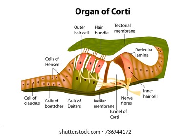
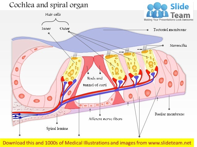
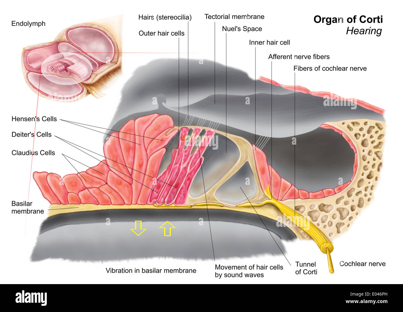
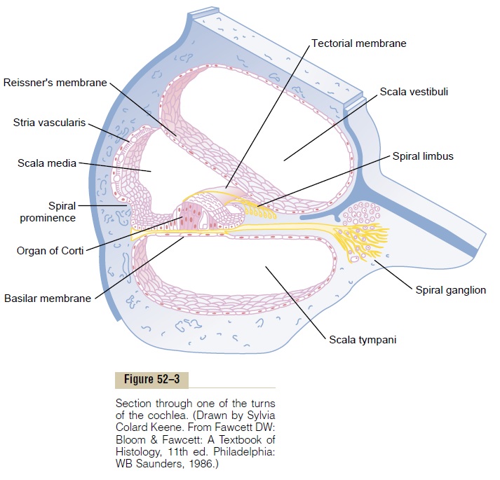
Schematic of the organ of Corti. In this transverse section of the basal part of a mammalian cochlea, 1 IHC (1) and 3 OHCs (2) are represented on either side of the tunnel of Corti (3). The tectorial membrane (6), floating in endolymph, caps the tallest stereocilia of the hair cells. The IHC is surrounded by supporting cells, whereas OHCs are ... In this video, we explore the cochlea and the Organ of Corti by viewing it under light microscopy.
Anatomy of the Ear. The ear is made up of three parts: the outer, middle, and inner ear. All three parts of the ear are important for detecting sound by working together to move sound from the outer part through the middle and into the inner part of the ear. Ears also help to maintain balance.

Organ of corti diagram
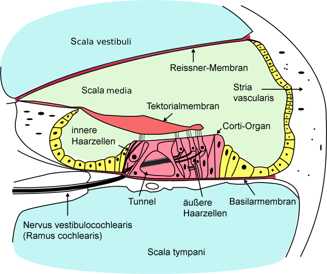
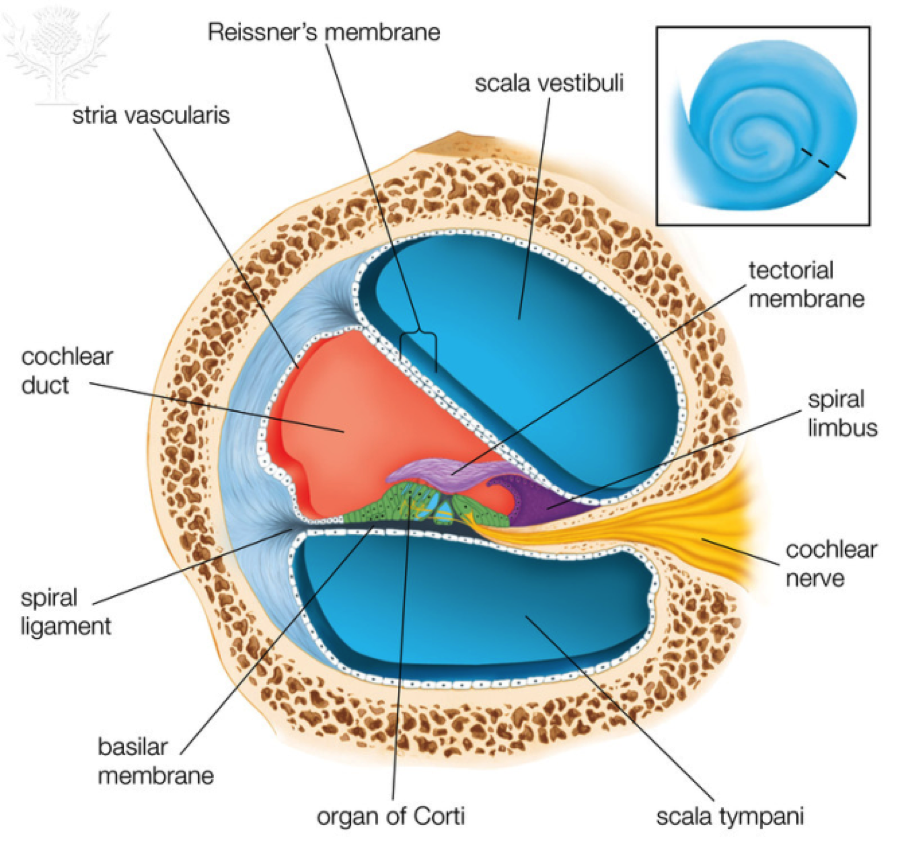
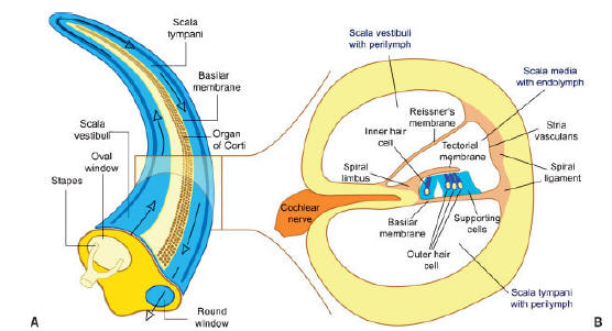
Organ Of Corti Diagram. organ of corti the organ of corti surrounded in potassium rich fluid endolymph lies on the basilar membrane at the base of the scala media under the organ of corti is the scala tympani and above it the scala vestibuli both structures exist in a low potassium fluid called perilymph diagram organ corti unity panies rr school c detail of the organ of corti hairs ... 13.03.2021 · The styloid process of the temporal bone is a slender osseous projection that points anteroinferiorly from the inferior surface of the petrous part of the temporal bone. It serves as an anchor point for several muscles associated with the tongue... The organ of Corti itself is located on the basilar membrane. The organ of Corti rests on the basilar membrane and contains two types of hair cells: inner hair cells and outer hair cells. Inner hair cells transduce sound from vibrations to neural signals via the shearing action of their stereocilia. Outer hair cells serve a function as acoustic ...
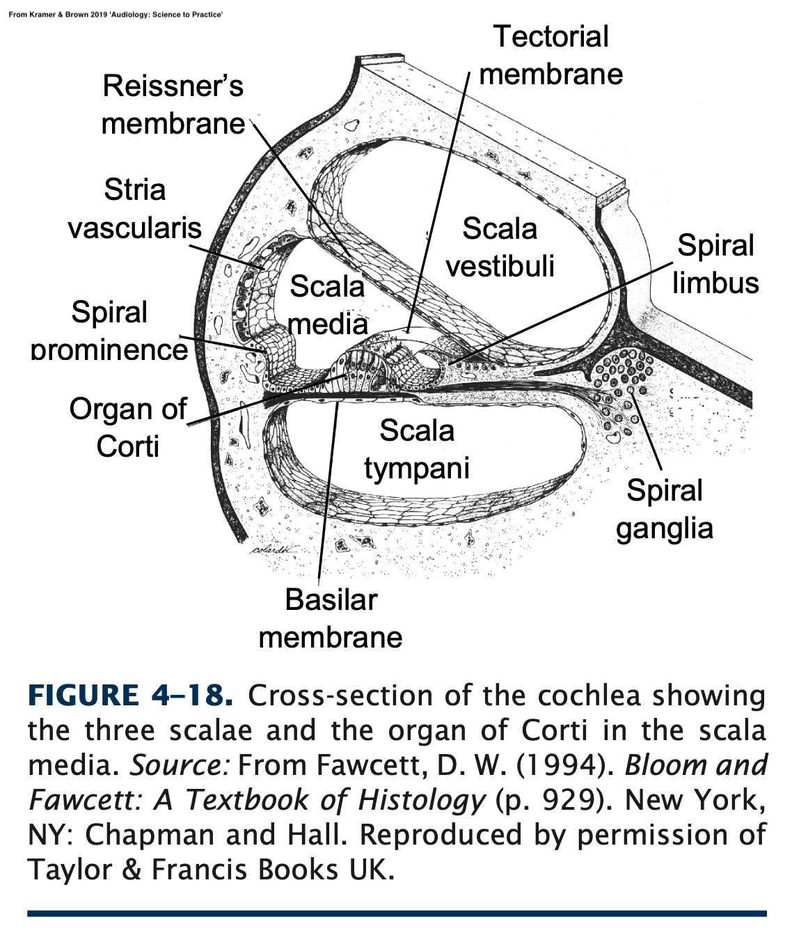
Organ of corti diagram. Diagram Of Organ Of Corti. The inner ear is housed in a maze of temporal bone passageways called the bony labyrinth, which is lined by a system of fleshy tubes called the membranous labyrinth (fig. 16.11). Between the bony and membranous labyrinths is a cushion of fluid called perilymph (PER-ih-limf), similar to cerebrospinal fluid. Perilymph is an extracellular fluid located within the inner ear.It is found within the scala tympani and scala vestibuli of the cochlea.The ionic composition of perilymph is comparable to that of plasma and cerebrospinal fluid.The major cation in perilymph is sodium, with the values of sodium and potassium concentration in the perilymph being 138 mM and 6.9 mM, respectively. DIAGRAM Diagram Of Organ Of Corti FULL Version HD Quality . Structure Organ Corti Stock Vector Royalty Free 736944172. Human earOrgan of CortiBritannica. Schematic representation of the organ of Corti. The figure . Organ of CortiPsychology WikiFandom. The Organ of Corti in the Inner Ear. Profitez de millions d'applications Android récentes, de jeux, de titres musicaux, de films, de séries, de livres, de magazines, et plus encore. À tout moment, où que vous soyez, sur tous vos appareils.
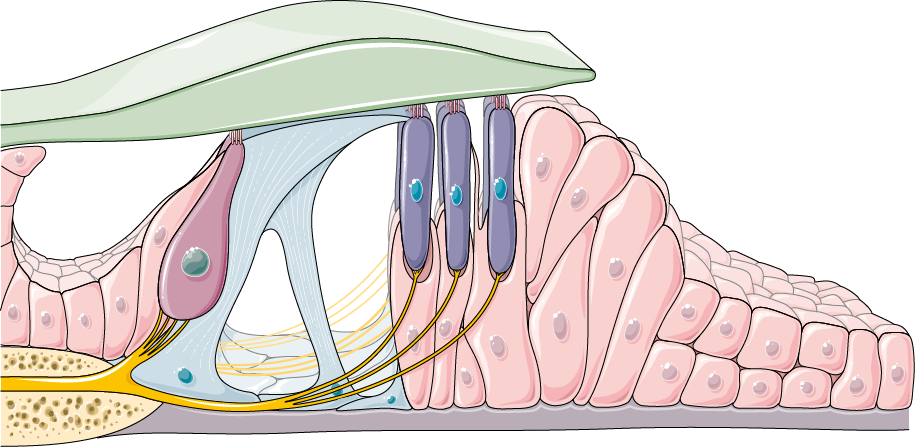
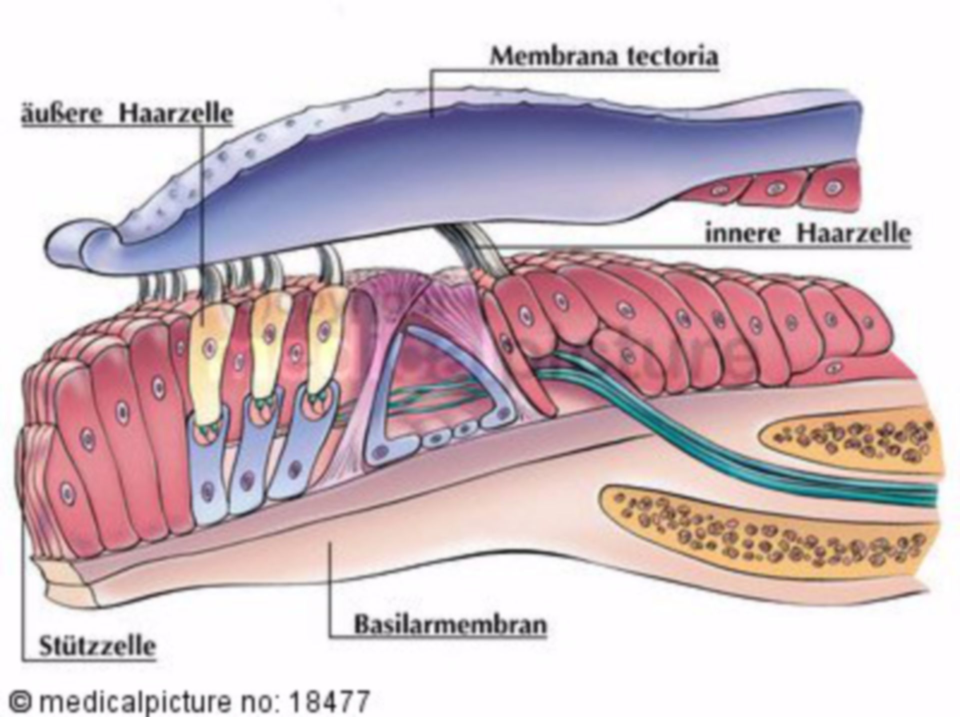
Schematic representation of the organ of Corti. The figure shows the different cell types and extracellular structures in the organ of Corti. In the diagram given below each triangle represents t h e c u s t o m e r s w h o have T.V. c h a n n e l connections. Which triangle(s) show the subscribers who have c o n n e c t i o n s of all t h e channels? (a) ZEE (b) SONY (c) STAR & UDAYA (d) ZEE & SONY ""BIOLOGY TODAY | MARCH '07 37. Which consonant among the given responses is common if three meaningful words are made from the given ... The organ of corti is a tube with hair-like cells that take the sound vibrations and changes them into electricity. Our brain runs on electricity; for it to understand sound, we have to change sound into electricity. When the hair-like cells are pushed by the sound, an important change happens. ... The Organ of Corti is an organ of the inner ear located within the cochlea which contributes to audition. The Organ of Corti includes three rows of outer hair cells and one row of inner hair cells. Vibrations caused by sound waves bend the stereocilia on these hair cells via an electromechanical force. The hair cells convert mechanical energy into electrical energy that is transmitted to the ...
The organ of Corti, or spiral organ, is the receptor organ for hearing and is located in the mammalian cochlea. This highly varied strip of epithelial cells ...Part of: Cochlea of the inner earNeuroLex ID: birnlex_2526Latin: organum spiraleStructure · Function · Development · Clinical significance The organ of Corti is the sensitive element in the inner ear and can be thought of as the body's microphone. It is situated on the basilar membrane in one of the three compartments of the Cochlea.It contains four rows of hair cells which protrude from its surface. Above them is the tectoral membrane which can move in response to pressure variations in the fluid- filled tympanic and vestibular ... …the basilar membrane is the organ of Corti, which contains the hair cells that give rise to nerve signals in response to sound vibrations. The side of the ... The organ of Corti itself is located on the basilar membrane. The organ of Corti rests on the basilar membrane and contains two types of hair cells: inner hair cells and outer hair cells. Inner hair cells transduce sound from vibrations to neural signals via the shearing action of their stereocilia. Outer hair cells serve a function as acoustic ...
Organ Of Corti Wikiwand
13.03.2021 · The styloid process of the temporal bone is a slender osseous projection that points anteroinferiorly from the inferior surface of the petrous part of the temporal bone. It serves as an anchor point for several muscles associated with the tongue...

The Ear Hair Cells Organ Of Corti The Auditory Nerve Video Lesson Transcript Study Com
Organ Of Corti Diagram. organ of corti the organ of corti surrounded in potassium rich fluid endolymph lies on the basilar membrane at the base of the scala media under the organ of corti is the scala tympani and above it the scala vestibuli both structures exist in a low potassium fluid called perilymph diagram organ corti unity panies rr school c detail of the organ of corti hairs ...

The Ear Book Chapter Iopscience

Organ Of Corti An Overview Sciencedirect Topics
The Organ Of Corti In The Inner Ear

Structure Of Organ Of Corti Download Scientific Diagram

Diagram Of Organ Of Corti Unity Companies Rr School Of Nursing
1

Spiral Organ Organ Of Corti Human Physiology 78 Steps Health
Structure Organ Corti Stock Vector Royalty Free 736944172
Where Is The Organ Of Corti Located Quora
Organ Of Corti Servier Medical Art

The Inner Ear Is Contains The Cochlea And The Auditory Nerve Description From Refinednotesblog Blogspot Com I Searc Ear Diagram Ear Anatomy Inner Ear Diagram

Inner Ear Diagram Outer Ear Hearing Ears Hand People Cartoon Png Pngwing
The Organ Of Corti Is Situated On The A Basilar Membrane Class 11 Biology Cbse

Cochlea And Organ Of Corti Open Educational Resource Oer Unsyiah Library

039 The Function Of The Organ Of Corti Youtube

What Is Organ Of Corti Biology Questions
Cochlea And Spiral Organ Medical Images For Power Point
Lign 113 Organ Of Corti Anatomy
Cochlea
Answers To This Module
The Organ Of Corti In The Inner Ear

Organ Of Corti In The Ear Stock Vector Illustration Of Tympani 60932666

Organ Of Corti Diagram Quizlet
Anatomy Of Cochlea
Shutterstock Puzzlepix
Why Is The Organ Of Corti An Organ Quora

Organ Of Corti Potentials And The Motion Of The Basilar Membrane Journal Of Neuroscience
1
Anatomy Of The Organ Of Corti Part Of The Cochlea Of The Inner Ear Stock Photo Alamy
Corti Organ Cochlea Doccheck

Auditory System Organ Of Corti
Function Of The Organ Of Corti

Organ Of Corti

Hearing 3 2 The Anatomy Of The Cochlea Openlearn Open University Sd329 1

Schematic Representation Of The Organ Of Corti The Figure Shows The Download Scientific Diagram

89 Organ Of Corti Photos And Premium High Res Pictures Getty Images

Organ Of Corti Png Images Pngegg
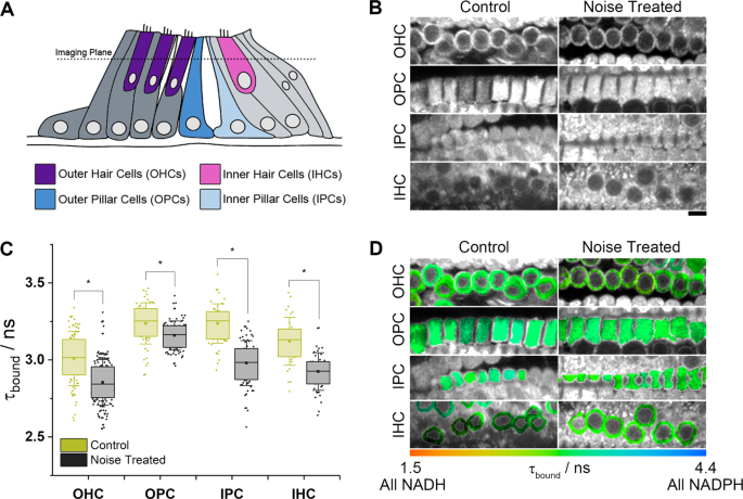
Multiphoton Nad P H Flim Reveals Metabolic Changes In Individual Cell Types Of The Intact Cochlea Upon Sensorineural Hearing Loss Scientific Reports
0 Response to "40 organ of corti diagram"
Post a Comment