39 labeled diagram of neuron
Parts of a Neuron Diagram. Although they have a characteristic elongated shape, they vary widely in size and properties based on their location and type of functions they perform. While they have the common features of a typical cell, they are structurally and functionally unique from other cells in many ways. component of nervous tissue neurons fall into types, illustration of labeled diagram of the neuron nerve cell that is the main part of the nervous system vector art Neuron Diagram Labeled 2020. Anatomical Organization Of Forward Fiber Projections From Area TE To » Picture Of A Neuron Labeled.
Neuron Labeled diagram of the Neuron, nerve cell that is the main part of the nervous system. Abstract grey mesh background. labeled diagram of a neuron stock illustrations Neural network vector illustration.
Labeled diagram of neuron
Neuron and Synapse Labeled Diagram. Human eye anatomy, retina, optic disc artery and vein etc. Structure of a motor neuron. Nerve damage. Human organs thin icons. Synapse and Neuron. Neuron types. Motor neuron. Vector diagram. Labeled diagram of the Neuron, nerve cell that is the main part of the nervous system. Abstract grey mesh background. Myelin sheath of the neuron. A schwann cell envelops and rotates around the axon forming myelin sheath, now axon is myelinated. Close-up detailed anatomy illustration; Sep 29, 2021 · Correia et al. show that muscle-secreted neurturin acts on muscle fibers and motor neurons to couple their characteristics in a functional way. This induces a shift toward a slow motor neuron identity and muscle oxidative metabolism and increases exercise performance and motor coordination in mice.
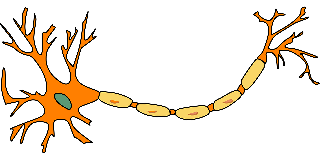
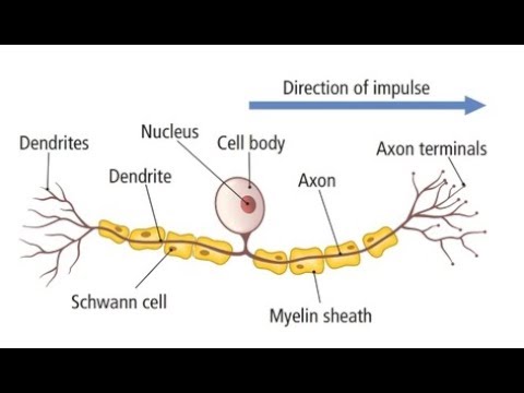
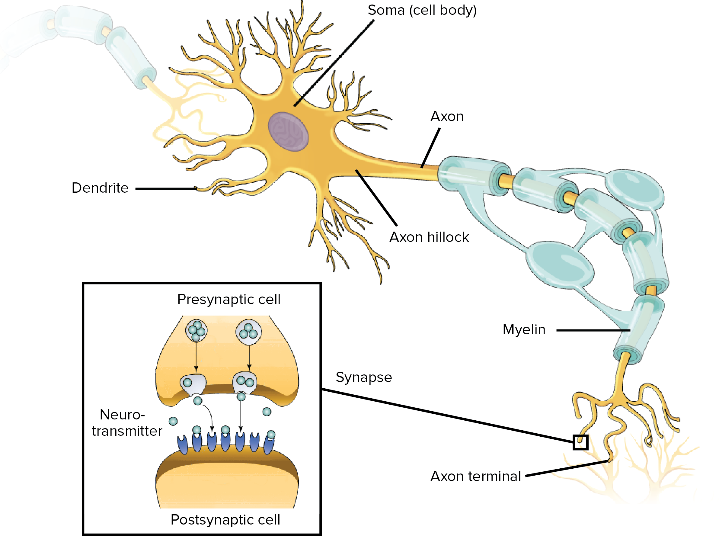
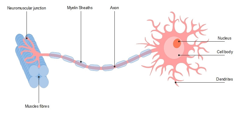
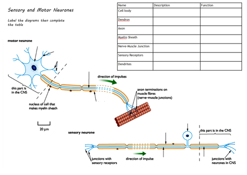
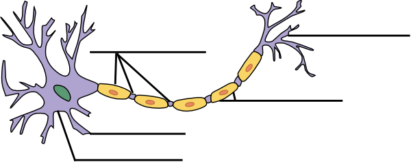
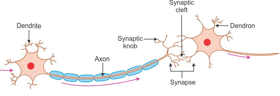
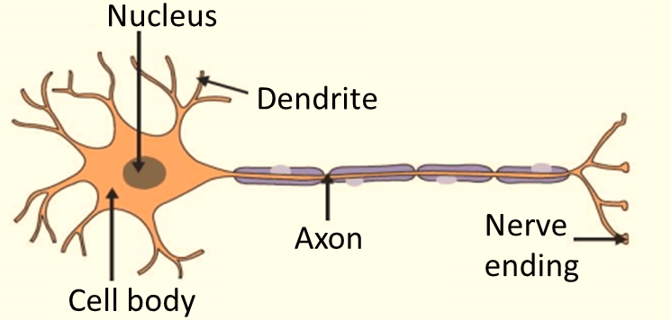
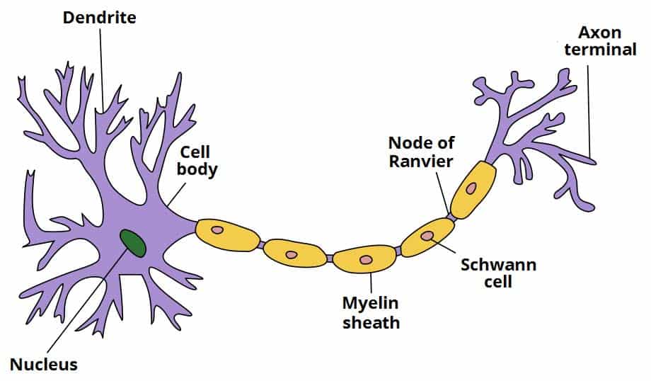
Labeled diagram of neuron. Draw a labelled diagram of a neuron Chapter 6: Tissue Science Class 9 solutions are avalable at ourwebsite to help the students. Q No 9: Draw a labelled diagram of a neuron. is solved by our expert teachers. You can get ncert solutions and notes for class 9 chapter 6 absolutely free. Diagram of Neuron. A neuron is a type of cell that is largely responsible for conveying information via electrical and chemical impulses. The brain, spinal cord, and peripheral nerves all contain them. The nerve cell is another name for a neuron. The structure of a neuron changes depending on its form and size, as well as its function and location. Neuron Anatomy Activity 1. Synapses: Send electrical impulses to neighboring neurons. 2. Myelin sheaths: Cover the axon and work like insulation to help keep electrical signals inside the cell, which allows them to move more quickly. 3. Axon: Transfers electrical impulse signals from the cell body to the synapse. 4. Diagram Of Neuron with Labels · Dendrites–A branch-like structure that functions by receiving messages from other neurons and allow the transmission of messages ...Cell Membrane Definition: Opening and Closing ...Placenta Definition: Introns and ExonsFermentation Definition: Meiosis DefinitionMitochondria Meaning: Rain Water Harvesting ...
Sep 18, 2021 · What is Artificial Neuron. An artificial neuron is a mathematical function based on a model of biological neurons, where each neuron takes inputs, weighs them separately, sums them up and passes this sum through a nonlinear function to produce output. In the next section, let us compare the biological neuron with the artificial neuron. Draw a labelled diagram of the neuron and describe the structure of the neuron in detail. · 1. Cell body: It forms the cytoplasm of the nerve cell. It is ...1 answer · Top answer: Hint: The cell forms the basic unit of life. Every part of the body has a specialized cell which will perform only a particular activity. They work ... Please watch: "cell structure and functions / animal cell vs plant cell / parts of cell / ch 8 science class 8 cbse" https://www.youtube.com/watch?v=surKjBAs... 3. How to Draw a Neuron Diagram To learn about the structure of the neurons, the students can use a neuron labeled diagram. The students may follow these steps to make their neuron diagram, but the process is complex: 3.1 How to Draw a Neuron Diagram from Sketch Step 1: First, the students need to draw a circle. Based on it, they need to draw a ...
Make a Neuron For grades 3-12. Create a model of a neuron by using clay, playdough, styrofoam, recyclables, food or anything else you can get your hands on. Use pictures from books to give you an idea of where the components of a neuron should go and what shape they should be. Use different colors to indicate different structures. Draw a labelled diagram of a neuron. Neurons are the fundamental unit of the nervous system.The neuron is a specialized and individual cell, which is also known as the nerve cell. A group of neurons forms a nerve. Dendrites - A branch-like structure that functions by receiving messages from other neurons and allow the transmission of messages ... A simple unlabeled tactile diagram of a neuron. Tactile Neuron Diagram ( Unlabeled) by trynne is licensed under the Creative Commons.Neuron Anatomy Activity The parts of the neuron have been labeled. Your challenge is to write the correct name for each part and explain what it does. neuron, (1). axon, cell body, dendrites, nucleus, terminal. Unlabeled diagram of a motor neuron (try labeling: axon, dendrite, cell body, myelin, nodes of Ranvier, motor end plate).Read the definitions, then label the neuron diagram below. axon - the long extension of a neuron that carries nerve impulses away from the body of the cell.
1
Neuron Unit Synapse Connection Synaptic strength Weight Firing frequency Signals pass fromUnit output Table 1 (left): Corresponding terms from biological and artificial neural networks. Adapted from Adapted from Mehrotra, Mohan, & Ranka. Figure 1 (below): Schematic diagram of a standard neural network design. the input units

File Neuron Or Nerve Cell Labelled Image Png Wikimedia Commons
Diagrams! They remind me of school textbooks which used to have plenty of them, providing a visual aid to understanding difficult subjects. This article explains the nervous system function and structure with the help of a human nervous system diagram and gives you that erstwhile 'textbook feel'. Read on.
Labeled Neuron Diagram Science Trends
06-10-2021 · Here we report the generation of a multimodal cell census and atlas of the mammalian primary motor cortex as the initial product of the BRAIN Initiative Cell …
How To Draw Sensory Neuron Well Labelled And Very Easy Youtube
Nodes of Ranvier: Location And Function (With a Labeled Diagram) Nodes of Ranvier are located between sheaths of myelin. They facilitate speedy electric transmission. Bodytomy, in this post, ... Each neuron in the network transmits the received impulse to the next neuron, until the required response is …

Draw A Labelled Diagram Of A Neuron Scholr
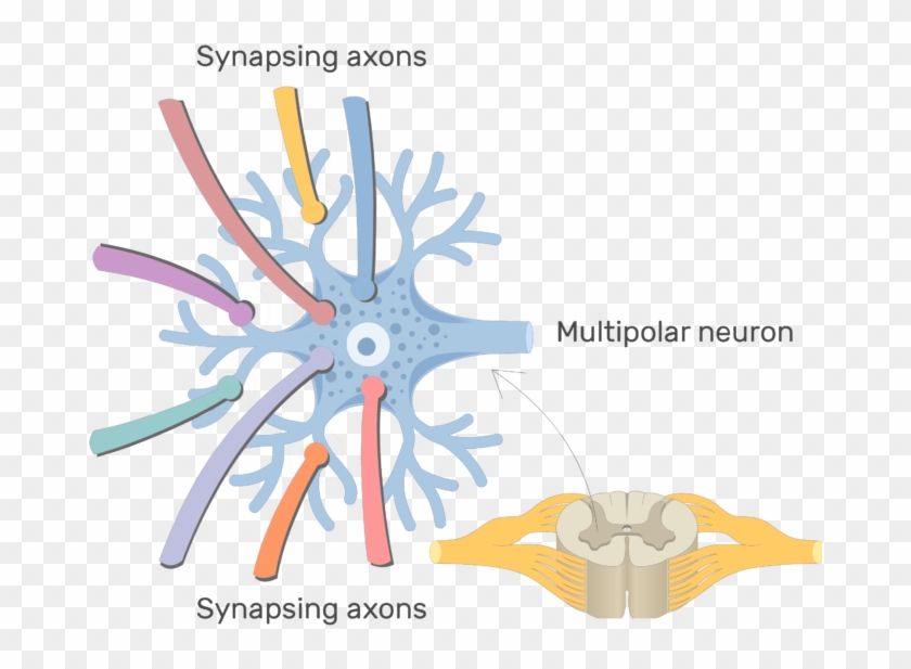
The neuron anatomy poster. Nervous system illustration. Different kinds of neurons: Bipolar neuron, Multipolar neuron, Unipolar neuron. Human nervous system medical vector illustration diagram with parasympathetic and sympathetic nerves and connected inner organs. Peripheral nervous system, medical vector illustration diagram with brain, spinal ...

Motor Neuron Alila Medical Images
Download the Diagram of Neuron Anatomy 358962 royalty-free Vector from Vecteezy for your project and explore over a million other vectors, icons and clipart graphics!

Types Of Neurons Vector Illustration Labeled Nerve Parts Comparison Scheme Stock Illustration Download Image Now Istock
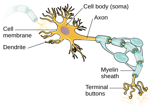
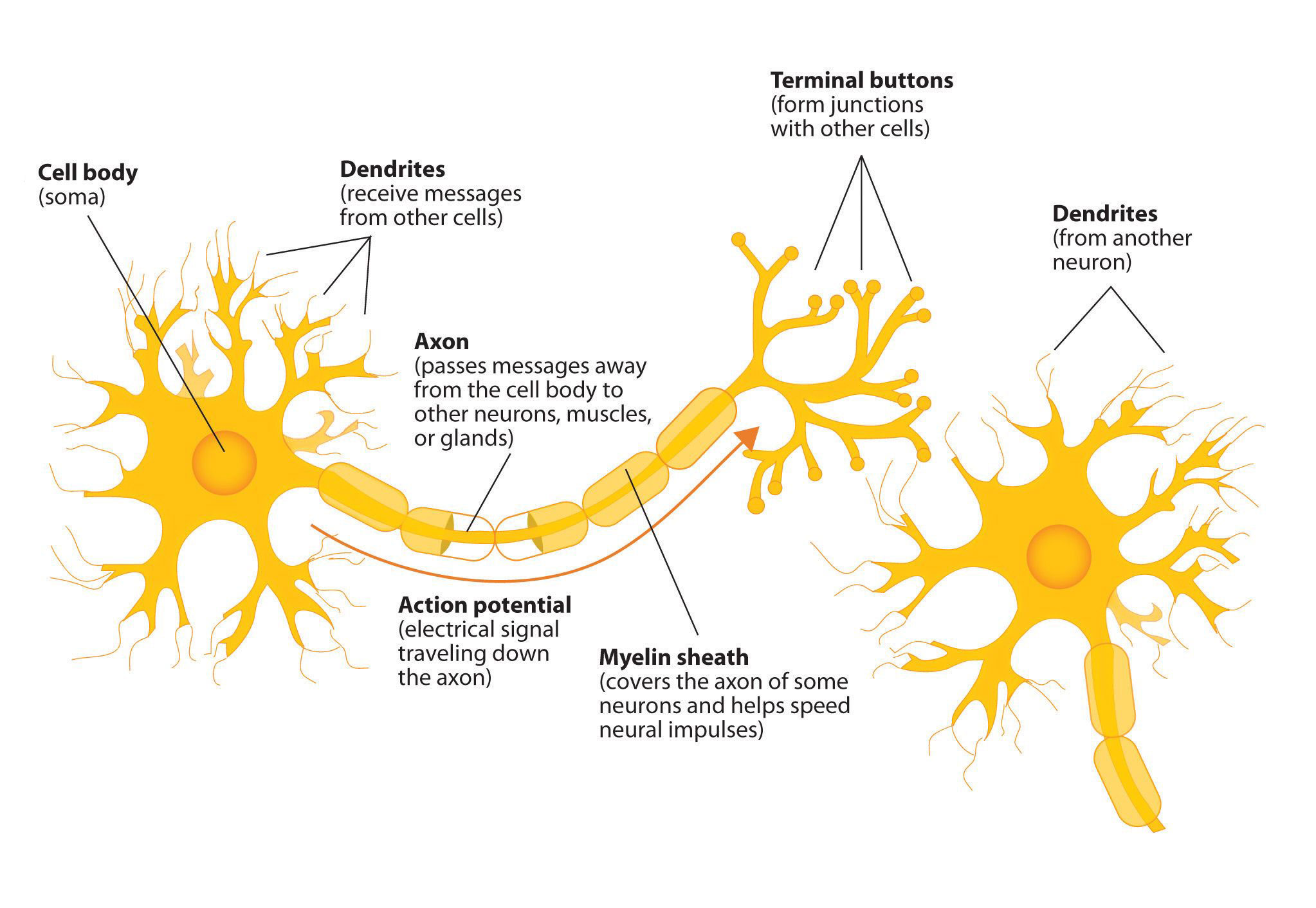
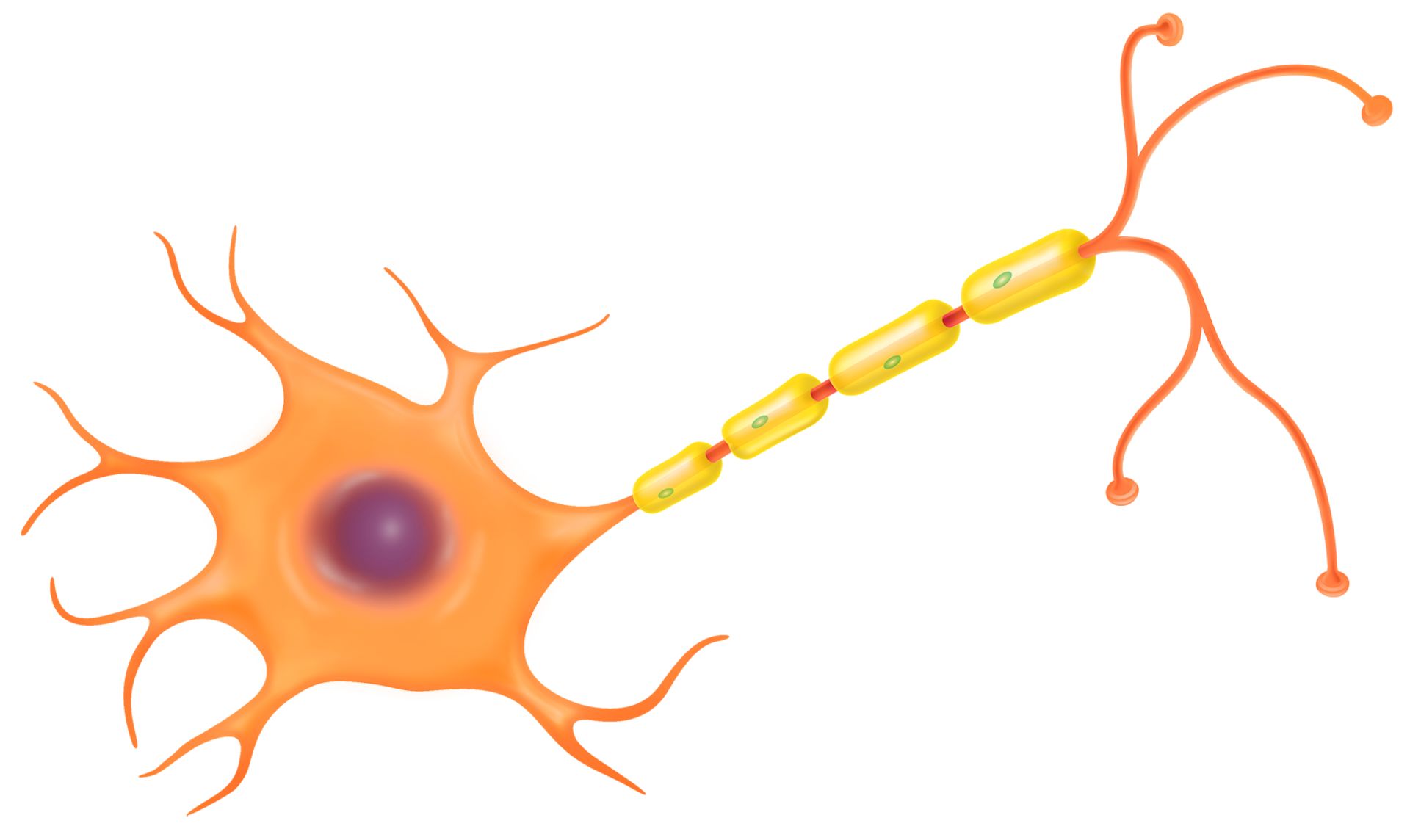
Anatomy of a neuron. Neurons, like other cells, have a cell body (called the soma ). The nucleus of the neuron is found in the soma. Neurons need to produce a lot of proteins, and most neuronal proteins are synthesized in the soma as well. Various processes (appendages or protrusions) extend from the cell body.
Overview Of Neuron Structure And Function Article Khan Academy
The epineurium is the outermost layer of dense irregular connective tissue surrounding a peripheral nerve. It usually surrounds multiple nerve fascicles as well as blood vessels which supply the nerve.
A Guide To Understand Neuron With Neuron Diagram Edrawmax Online
Labeled Neuron Diagram. Alex Bolano on May 29, ... The human brain is estimated to contain over 100 billion neurons with each neuron having, on average, connections to 1000 other neurons. This vast connectional architecture explains that computational complexity of the human brain. Neurons communicate with each other by generating and ...

Labeled Diagram Of The Neuron Canvas Print Barewalls Posters Prints Bwc31569020
Nervous System - Neuron: Nerve Cell. Choose the correct names for the parts of the neuron. This neuron part receives messages from other neurons. This neuron part sends on messages to other neurons. This neuron part gives messages to muscle tissue. This neuron part processes incoming messages.
Nervous System Structure Function And Diagram Kenhub
A neuron or nerve cell is an electrically excitable cell that communicates with other cells via specialized connections called synapses.It is the main component of nervous tissue in all animals except sponges and placozoa. Plants and fungi do not have nerve cells.. Neurons are typically classified into three types based on their function. Sensory neurons respond to stimuli such as touch, sound ...

Draw A Labelled Diagram Of A Neuron Science Shaalaa Com
Start studying Label Parts of a Neuron. Learn vocabulary, terms, and more with flashcards, games, and other study tools.
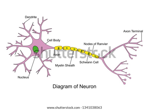
Simple Neuron Diagram 2d Labeled Nerve Stock Vector Royalty Free 1341038063
May 15, 2021 · Animal cell size and shape. Animal cells come in all kinds of shapes and sizes, with their size ranging from a few millimeters to micrometers. The largest animal cell is the ostrich egg which has a 5-inch diameter, weighing about 1.2-1.4 kg and the smallest animal cells are the neurons of about 100 microns in diameter.

Draw A Labeled Diagram Of A Neuron Cbse Master Ncert Textbooks Exercises Solutions
Neuron Anatomy. Nerve Cell: Dendrites receive messages from other neurons. The message then moves through the axon to the other end of the neuron, then to the tips of the axon and then into the space between neurons. From there the message can move to the next neuron. Neurons pass messages to each other using a special type of electrical signal.

34 Drawing Of Neuron And Label Of Its Part Labels Database 2020
Cell Labeling Chart. This is a free printable chart of the animal cell featuring each of the different parts labeled for children to learn. This is a great resource for hanging in your classroom or adding to your science notebook
Neuron Labeled Images Stock Photos Vectors Shutterstock
Draw a labelled diagram of the neuron and describe the structure of neuron in detail. Medium. Open in App. Solution. Verified by Toppr. The structure of neuron: Nerve cells or neurons are the structural and functional units of the nervous system. It consists of three major parts namely, Cell body, dendrites, Axon.
Draw A Labelled Diagram Of A Myelinated Neuron Sarthaks Econnect Largest Online Education Community
labeled diagram of nerve cell · Asked by sgg1400 | 8th Apr, 2018, 09:29: AM · Structure of a nerve cell: · Answered by Sheetal Kolte | 9th Apr, 2018, ...

Bmsc11001 Label Diagram Neuron Diagram Quizlet
English: Complete neuron cell diagram. Neurons (also known as neurones and nerve cells) are electrically excitable cells in the nervous system that process and transmit information. In vertebrate animals, neurons are the core components of the brain, spinal cord and peripheral nerves. Own work.
Neurons Introduction To Psychology
This is an online quiz called Label a Neuron. There is a printable worksheet available for download here so you can take the quiz with pen and paper. From the quiz author. Title says it all. Your Skills & Rank. Total Points. 0. Get started! Today's Rank--0. Today 's Points. One of us! Game Points. 10.
Neuron Clipart Generic Labeled Multipolar Neuron Diagram Hd Png Download 694x550 6620046 Pngfind
Find neuron labeled stock images in HD and millions of other royalty-free stock ... Neuron and Synapse Labeled Diagram ... Nerve Cell Neuron Labeled Diagram.
Neurone Diagrams To Label Teaching Resources
Illustration of Labeled diagram of the neuron, nerve cell that is the main part of the nervous system. vector art, clipart and stock vectors.
Label The Neuron Clip Art At Clker Com Vector Clip Art Online Royalty Free Public Domain
Rubrospinal tract is labeled in red on the left of the diagram. Schematic representation of the chief ganglionic categories (Rubrospinal tract not labeled, but red nucleus visible near center) Details

Neuron Diagram Stock Illustration Download Image Now Istock
Structure. The white rami communicantes are the preganglionic sympathetic outflow from the spinal cord. The cell bodies for the preganglionic sympathetic myelinated fibers in the white rami communicantes lie in the ipsilateral (same sided) intermediolateral cell column in the spinal cord which extends from T1-L2.
Describe The Microscopic Structure Of A Neuron Also Draw Its Diagram Studyrankersonline
Start studying neuron labeled. Learn vocabulary, terms, and more with flashcards, games, and other study tools.

Neuron And Synapse Labeled Diagram Royalty Free Cliparts Vectors And Stock Illustration Image 31325122
Mar 04, 2021 · Cell membranes protect and organize cells. Which best Describes How the function of the Cell Membrane differs in plant and animal Cells A) In Animal Cells the cell Membrane is the outermost layer of the cell while in plant cells, the cell membrane separates the cell wall from everything inside the the cell The animal cell is made up of several structural organelles enclosed in the plasma ...

How To Draw Structure Of Neuron Neuron Diagram Labelled Diagram Of Neuron Neuron Cell Youtube Cell Diagram Neuron Diagram Nerve Cell
Click here to get an answer to your question ✍️ Draw neat labelled diagram of neuron.

Diagram Quiz On Neuron Structure And Function Labeling Quiz
Draw a labeled diagram of neuron. 2 2. Write one term for the following: a) joins muscle to bone b) fat reservoir of our body c) supporting, fills the space inside the organs and help in repair of tissues 3 3. Label the diagram and give one function each of a,b and c 3 4.

Draw A Labelled Diagram Of Neuron And Label Any Four Different Parts Explain Brainly In
Labeled diagram of the neuron, nerve cell that is the main part of the nervous system. Anatomy of a nerve, eps8; Neuron, nerve cell, close up illustrations set. Synapse detailed anatomy, neuron passes signal to another neuron. Cross section, nucleus and other organelles of the cell.

Draw A Labelled Diagram Of A Neuron Brainly In
100% (2 ratings) Multipolar motor neurons are found in central nervous system (CNS). Multipolar motor neurons contain multiple dendrite …. View the full answer. Transcribed image text: ures Nucieus Axon FIQURE 25.1 Label this diagram of a mutipolar motor neuron BPR.
Draw A Diagram Of Neuron And Name And Label The Part A Where Information Is Acquired B Through Which Information Travels As An Electric Impulse And C Where The Electric Impulse Must
Sep 29, 2021 · Correia et al. show that muscle-secreted neurturin acts on muscle fibers and motor neurons to couple their characteristics in a functional way. This induces a shift toward a slow motor neuron identity and muscle oxidative metabolism and increases exercise performance and motor coordination in mice.

Explain The Structure Of Neuron With The Help Of A Labelled Diagram Science Shaalaa Com
Labeled diagram of the Neuron, nerve cell that is the main part of the nervous system. Abstract grey mesh background. Myelin sheath of the neuron. A schwann cell envelops and rotates around the axon forming myelin sheath, now axon is myelinated. Close-up detailed anatomy illustration;

Labeled Diagram Of The Neuron
Neuron and Synapse Labeled Diagram. Human eye anatomy, retina, optic disc artery and vein etc. Structure of a motor neuron. Nerve damage. Human organs thin icons. Synapse and Neuron. Neuron types. Motor neuron. Vector diagram.

Label Neuron Anatomy Printout Enchantedlearning Com

Draw A Labelled Diagram Of A Neuron
Very Short Answer Question Draw A Well Labelled Diagram Of Neuron Cell Snapsolve
Ultrastructure Of Nerves Classification Neurones Teachmeanatomy
Neuron Labeled Diagram Anatomy And Structure
Nerve Cell Function Nerve Cell Diagram Dk Find Out

Parts Of A Neuron Diagram Quizlet
0 Response to "39 labeled diagram of neuron"
Post a Comment