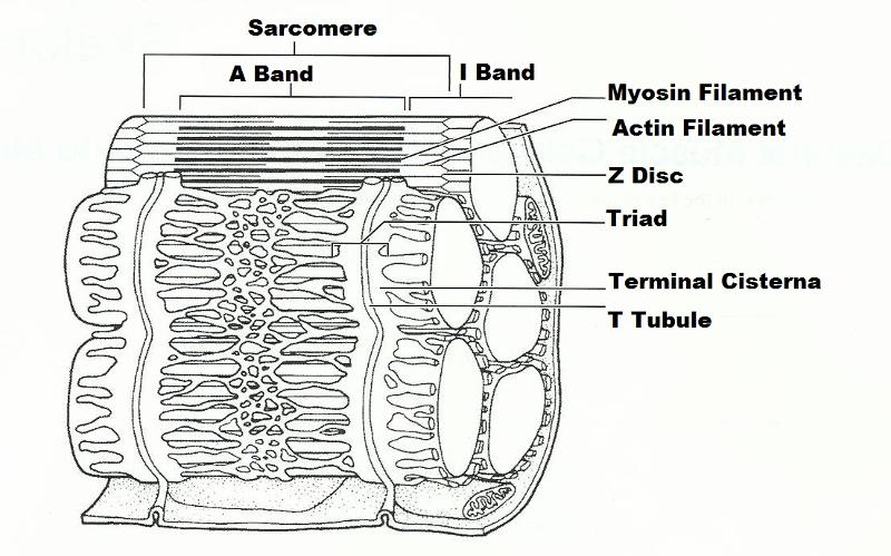
38 the diagram illustrates a small portion of several myofibrils
The diagram illustrates a small portion of several myofibrils. Using letters from the key, correctly identify each structure.3 pages There are several reasons for this. First. Show what is diagram illustrates of several myofibrils? . The function of a myofibril in short is to shorten and to contract. Show what Diagrams to illustrate and explain the impact on the equilibrium wage rate and quantity of labor supplied in.The diagram illustrates a small portion of several myofibrils.
The diagram below provides the platform for connection that the business employs to connect farmers with their respective clients. Part B: customers access Adopt-A-Farm website to get information. ... the diagram illustrates a small portion of several myofibrils; Cite this document. APA; MLA;
The diagram illustrates a small portion of several myofibrils
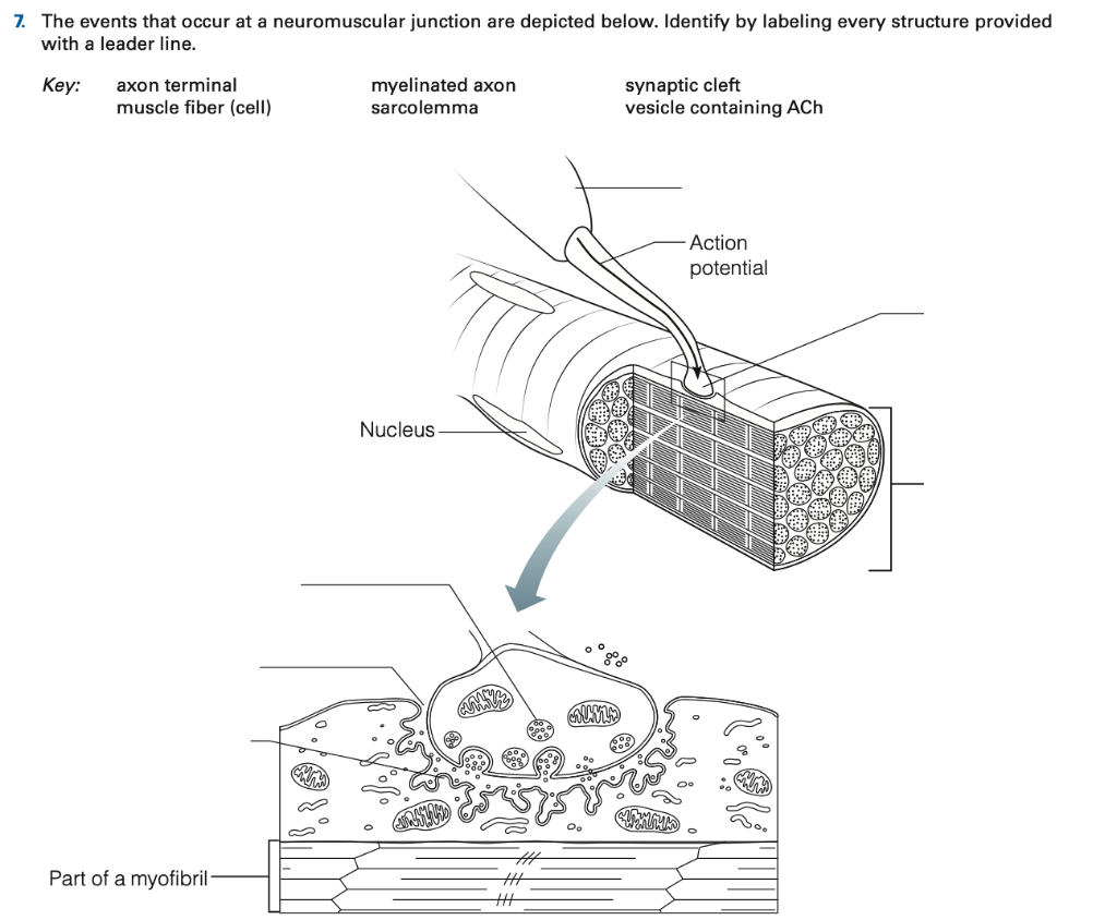
5. Figure 6—3 is a diagrammatic representation of a small portion of a relaxed muscle cell (bracket indicates the portion enlarged). First, select different colors for the structures listed below. Use them to color the coding circles and corre- sponding structures on Figure 6—3. Then bracket and label an A band, an I band, and a sarcomere. The Diagram Illustrates A Small Portion Of Several Myofibrils; Autopage Rs 730 Wiring Diagram; Comelit Intercom Wiring Diagram; Pioneer Avh P5700dvd Wiring Diagram; 2004 Grand Prix Monsoon Wiring Diagram; Xylem Diagrams; Delco Remy 10dn Wiring Diagram; Fender Jaguar Baritone Wiring Diagram; 4.3 Vortec Vacuum Diagram; Winco Generator Wiring Diagram Diagram & EM of NMJ Stuctures to identify (Mitochondria, Synaptic Vesicles, Junctional Folds (Red Arrow), Primary & Secondary synaptic clefts, Mitochondria, Nucleus of Muscle Fiber, Myofibrils) ***See Ross Histology pg 292 for details
The diagram illustrates a small portion of several myofibrils. 300zx Intercooler Piping Diagram; 164d3871p001 Parts Diagram; 2002 Chrysler Voyager 2.4 Engine Wiring Diagram; Argo V693-36 Wiring Diagram; Comets And Asteroids Venn Diagram; Rd200 Wiring Diagram; The Diagram Illustrates A Small Portion Of Several Myofibrils; Wiring Diagram Angled 3 Way Switchcraft; R30-1 Plug Wiring Diagram; 2006 Bmw 330i ... The mitochondria in muscle fiber are arranged: a. randomly b. in circles c. around the nuclei d. in rows close to the myofibrils throughout the muscle fiber e. closest to the sarcolemma d This part of the skeletal muscle fibert releases calcium when stimulated by the T tubules: a. myofibrils b. sarcoplasm c.sarcomeres d. terminal cisterna of ... Access Human Anatomy & Physiology Laboratory Manual, Fetal Pig Version, Update 10th Edition Chapter E14 Problem 5E solution now. Our solutions are written by Chegg experts so you can be assured of the highest quality! An aponeurosis is a sheet of white fibrous connective tissue; The diagram illustrates a small portion of several myofibrils. Using letters from the key, correctly identify each structure indicated by a leader line or a bracket. Key: a. b. c. A band actin filament I band d. e. f. myosin filament T tubule terminal cisterna g. h. i. triad ...
The The diagram illustrates a small portion of several myofibrils is one of the most popular assignments among students' documents. If you are stuck with writing or missing ideas, scroll down and find inspiration in the best samples. The diagram illustrates a small portion of several myofibrils is quite a rare and popular topic for writing an essay, but it certainly is in our database. s Dependency diagram and its use A dependency diagram is a graphical representation of a dependency chart graphs. These diagrams are vital in software pack development by outlining the complexity and the interrelationships of various functional elements (Bagui and Earp 739). However, in the dependency diagrams arrows point from the modules they ... The diagram illustrates a small portion of several myofibrils. Using letters from the key, correctly identify each structure indicated by a leader line or bracket. Key: a. A band d. myosin filament g. triad b. Actin filament e. T tubule h. sarcomere c. I band f. terminal cistern i. Z dis C I D A E H G F B Start studying a small portion of a muscle myofibril. Learn vocabulary, terms, and more with flashcards, games, and other study tools.
1. The diagram shows part of a muscle myofibril (a) Name the protein present in the filaments labelled W and X W = myosin, X = actin (b) Figure 2 shows the cut ends of the protein filaments when the myofibril was cut at position Y. figure 3 shows the protein filaments when the myofibril was cut at the same have knowledge that, people have look numerous time for their favorite books taking into ... The Diagram Illustrates A Small Portion Of Several Myofibrils. Access Human Anatomy Laboratory Manual with Cat Dissections 8th Edition Chapter E11 Problem 5E solution now. Our solutions are written by Chegg experts so you can be assured of the highest quality! Myofibril Definition. A myofibril is a component of the animal skeletal muscle. Myofibrils are long filaments that run parallel to each other to form muscle (myo) fibers. The myofibrils, and resulting myofibers, may be several centimeters in length. The muscle fibers are single multinucleated cells that combine to form the muscle.
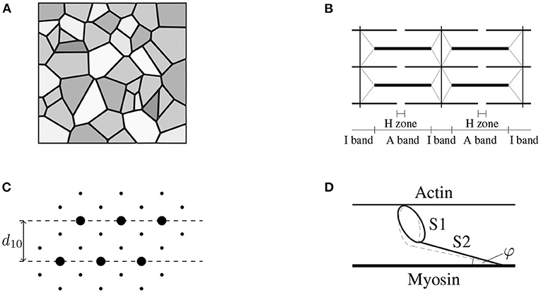
Ijms Free Full Text X Ray Diffraction Studies On The Structural Origin Of Dynamic Tension Recovery Following Ramp Shaped Releases In High Ca Rigor Muscle Fibers Html
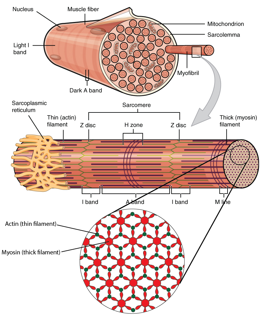
Figure 10.2.2 - Muscle Fiber: A skeletal muscle fiber is surrounded by a plasma membrane called the sarcolemma, which contains sarcoplasm, the cytoplasm of muscle cells. A muscle fiber is composed of many myofibrils, which contain sarcomeres with light and dark regions that give the cell its striated appearance.
Royalsocietypublishing Org
The diagram illustrates a small portion of a muscle myofibril in a highly simplified way. Using terms from the key, correctly.6 pages

Elucidation Of Thioredoxin Target Protein Networks In Mouse Molecular Cellular Proteomics
The diagram illustrates a small portion of a muscle myofibril. Using letters from the key ... Several criteria were given relative to the naming of muscles.

Ch 11 Lab Manual Pdf E X E R C Is E 11 Review Sheet Microscopic Anatomy And Organization Of Skeletal Muscle Name Lab Time Date Skeletal Muscle Cells Course Hero
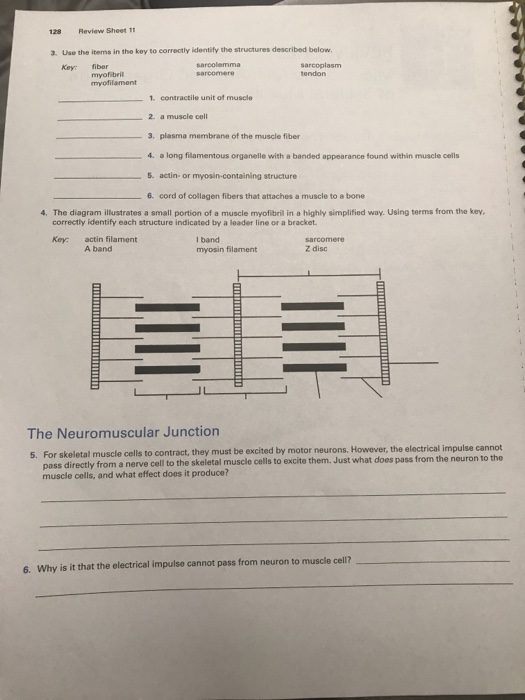
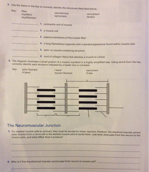
3. The diagram illustrates a small portion of several myofibrils. Using letters from the key, correctly identify each structure indicated by a leader line or bracket. Key: a. A band d. myosin filament g. triad b. Actin filament e. T tubule h. sarcomere c. I band f. terminal cistern i. Z disc
Skeletal Muscle Organization
But, this does not illustrate the complexities of the usage of the English language in India. ... the diagram illustrates a small portion of several myofibrils; Cite this document ... So it is to be understood in this paper that writing is an assumed literacy practice that is said to be an essential part of the performance, although it is not a ...
Three Distinct Sarcomeric Patterns Of Skeletal Muscle Revealed By Shg And Tpef Microscopy
The diagram illustrates a small portion of several myofibrils. Using letters from the key, correctly identify each ... To plot a graph relating stimulus strength and twitch force to illustrate graded muscle response. 4. To explain how slow, smooth, sustained contraction is possible in a skeletal muscle. ... Skeletal muscle is composed of ...
Pcsd Org
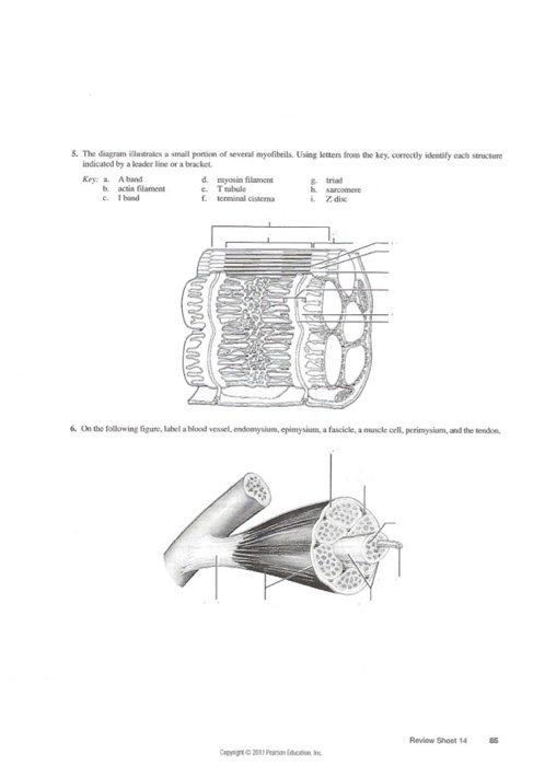
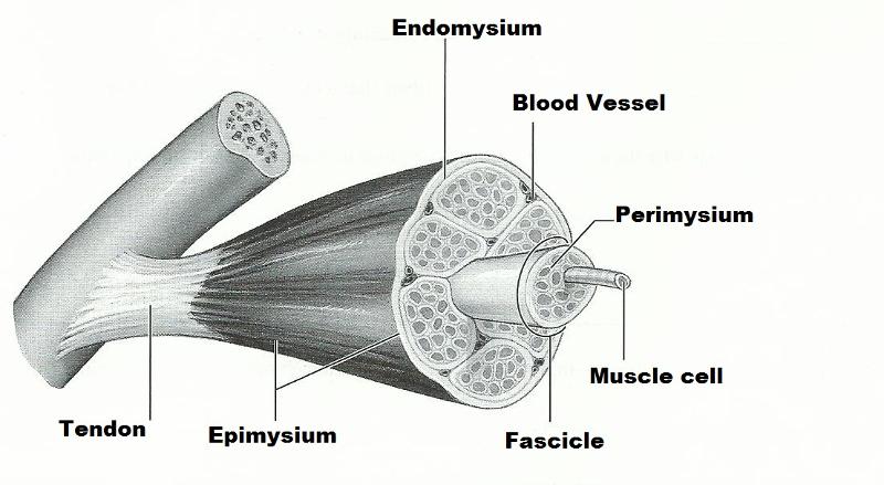
The diagram illustrates a small portion of several myofibrils. Correctly label all the structures defined in your lab manual. 2. On the following figure, label a blood vessel, endomysium, epimysium, a fasicle, a muscle cell, perimysium and the tendon.
Solved 4 The Diagram Illustrates A Small Portion Of A Chegg Com
The diagram illustrates a small portion of several myofibrils. Using letters from the key, correctly identify each structure indicated by a leader line or a bracket. Key: a. A band d. myosin filament g. triad b. actin filament e. T tubule h. sarcomere c. I band f. terminal cistern i. Z disc 6.
Ijms Free Full Text X Ray Diffraction Studies On The Structural Origin Of Dynamic Tension Recovery Following Ramp Shaped Releases In High Ca Rigor Muscle Fibers Html
The diagram illustrates a small portion of several myofibrils. Correctly label all the structures defined in your lab manual. Problem 5E: The diagram illustrates a small portion of several myofibrils. Using letters from the key, correctly identify each structure indicated by a leader line or a diagramweb.net:a. A bandb. actin filamentc.
Exercise 14 Microscopic Anatomy And Organization Of Skeletal Muscle Flashcards Easy Notecards
The diagram illustrates a small portion of several myofibrils. Using letters from the key, correctly identify each structure indicated by a leader line or a bracket. Key: a. a band d. myosin filament g. triad b. actin filament e. T tubule h. sarcomere c. I band f. terminal cistern i.
Solved Muscles Of The Head And Neck Using Choices From There Chegg Com
The Diagram Illustrates A Small Portion Of Several Myofibrils; Wiring Diagram For Kioti Dk45se; Pioneer Deh 1600 Wiring Diagram; Avh-601ex Wiring Diagram; Kao 5 Switch Wiring Diagram; Dual Xdm7510 Wiring Diagram; Wiring Diagram Xt2 Cub Cadet; Poulan Pro 260 Fuel Line Diagram; Servo Motor Wiring Diagram Adafruit; Autometer Phantom Tach Wiring ...

Schematic Diagram Illustrating The Major Sarcomeric Components Of Download Scientific Diagram
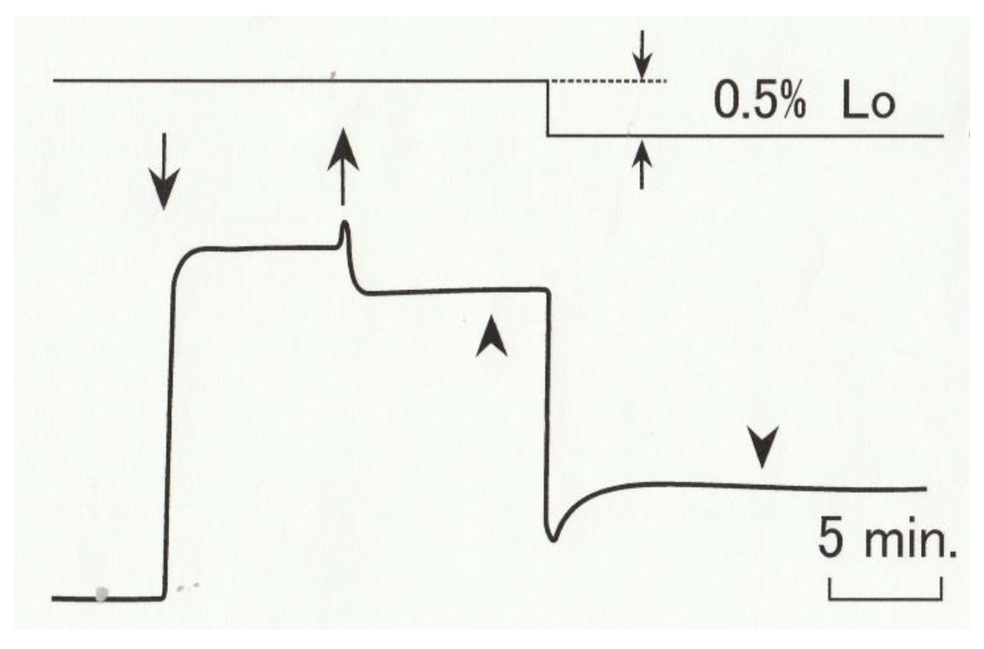
The diagram illustrates the single isometric twitch characteristics of two skeletal muscles, A and B, in response to a depolarizing stimulus. The delay between the termination of the transient depolarization of the muscle membrane and the onset of muscle contraction observed in both muscles A and B reflects the time necessary for which of the ...
Myofibrils High Resolution Stock Photography And Images Alamy
The multiple oval nuclei can be seen just beneath the plasma ... The diagram illustrates a small portion of a muscle myofibril in a highly simplified way.10 pages

Sarcoplasm An Overview Sciencedirect Topics
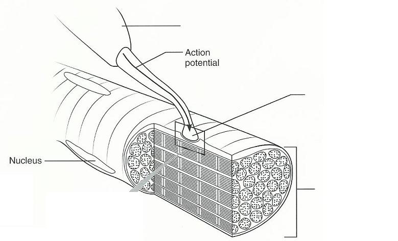
The diagram illustrates a small portion of several myofibrils. ... Within the axon terminal are many small vesicles containing a neurotransmitter substance ...
Exercise 14 Microscopic Anatomy And Organization Of Skeletal Muscle Flashcards Easy Notecards
The diagram illustrates a small portion of several myofibrils. Using letters from the key, correctly identify each structure indicated by a leader line or a bracket. Key: a A band d. myosin filament g triad b. actin filament e. Ttubule c. I band h. sarcomere t. terninal cisterm i. Zdisc 6. On the following figure, label a blood vessel,

18 3 The Molecular Structure And Sub Cellular Organization Of Cytoskeletal Components Biology Libretexts
Transcribed image text: Review sheet 14 Diagram illustrates a small portion of several myofibrils. Using letters from the key, correctly uted by a leader ...

References In A Rational Approach To Fluid Therapy In Sepsis British Journal Of Anaesthesia
Thus, PortionPac's business practice is therefore, highly recommendable. Answer 2 The most important and influential stakeholders for Portion Pac are employees who make them and the end users of its products which are designed for cleaning with least use of chemicals. Its end users are janitors, housekeepers and food service professionals.

Ch 11 Lab Manual Pdf E X E R C Is E 11 Review Sheet Microscopic Anatomy And Organization Of Skeletal Muscle Name Lab Time Date Skeletal Muscle Cells Course Hero
Diagram & EM of NMJ Stuctures to identify (Mitochondria, Synaptic Vesicles, Junctional Folds (Red Arrow), Primary & Secondary synaptic clefts, Mitochondria, Nucleus of Muscle Fiber, Myofibrils) ***See Ross Histology pg 292 for details
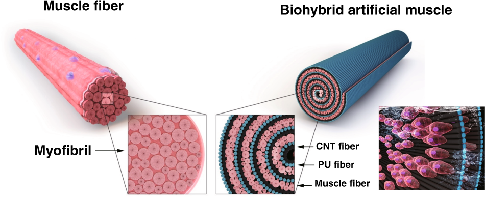
Biomimetic Cell Actuated Artificial Muscle With Nanofibrous Bundles Microsystems Nanoengineering
The Diagram Illustrates A Small Portion Of Several Myofibrils; Autopage Rs 730 Wiring Diagram; Comelit Intercom Wiring Diagram; Pioneer Avh P5700dvd Wiring Diagram; 2004 Grand Prix Monsoon Wiring Diagram; Xylem Diagrams; Delco Remy 10dn Wiring Diagram; Fender Jaguar Baritone Wiring Diagram; 4.3 Vortec Vacuum Diagram; Winco Generator Wiring Diagram

Review Sheet 11
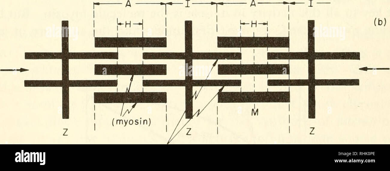
5. Figure 6—3 is a diagrammatic representation of a small portion of a relaxed muscle cell (bracket indicates the portion enlarged). First, select different colors for the structures listed below. Use them to color the coding circles and corre- sponding structures on Figure 6—3. Then bracket and label an A band, an I band, and a sarcomere.

Hypercontractile Properties Of Cardiac Muscle Fibers In A Knock In Mouse Model Of Cardiac Myosin Binding Protein C Journal Of Biological Chemistry

The Architectural Design Of The Gluteal Muscle Group Implications For Movement And Rehabilitation Journal Of Orthopaedic Sports Physical Therapy

Etiology Biology And Treatment Of Muscular Lesions Intechopen

Skeletal Muscle Cell An Overview Sciencedirect Topics
Solved 4 The Diagram Illustrates A Small Portion Of A Chegg Com
Frontiers A Physiology Guided Classification Of Active Stress And Active Strain Approaches For Continuum Mechanical Modeling Of Skeletal Muscle Tissue Physiology

Biology And Evolution Of The Mollusca
Core Ac Uk
Arxiv Org
Exercise 14 Microscopic Anatomy And Organization Of Skeletal Muscle Flashcards Easy Notecards
Solved 128 Review Sheet 11 3 Use The Items In The Key To Chegg Com

Jaypeedigital Ebook Reader
Solved Please Help Me Answer These Correctly And Completely Chegg Com

Marasmus Background Pathophysiology Body Composition

Phosphorylation In Novel Mitochondrial Creatine Kinase Tyrosine Residues Render Cardioprotection Against Hypoxia Reoxygenation Injury
Images Pcmac Org

Myofibril Diameter Is Set By A Finely Tuned Mechanism Of Protein Oligomerization In Drosophila Elife
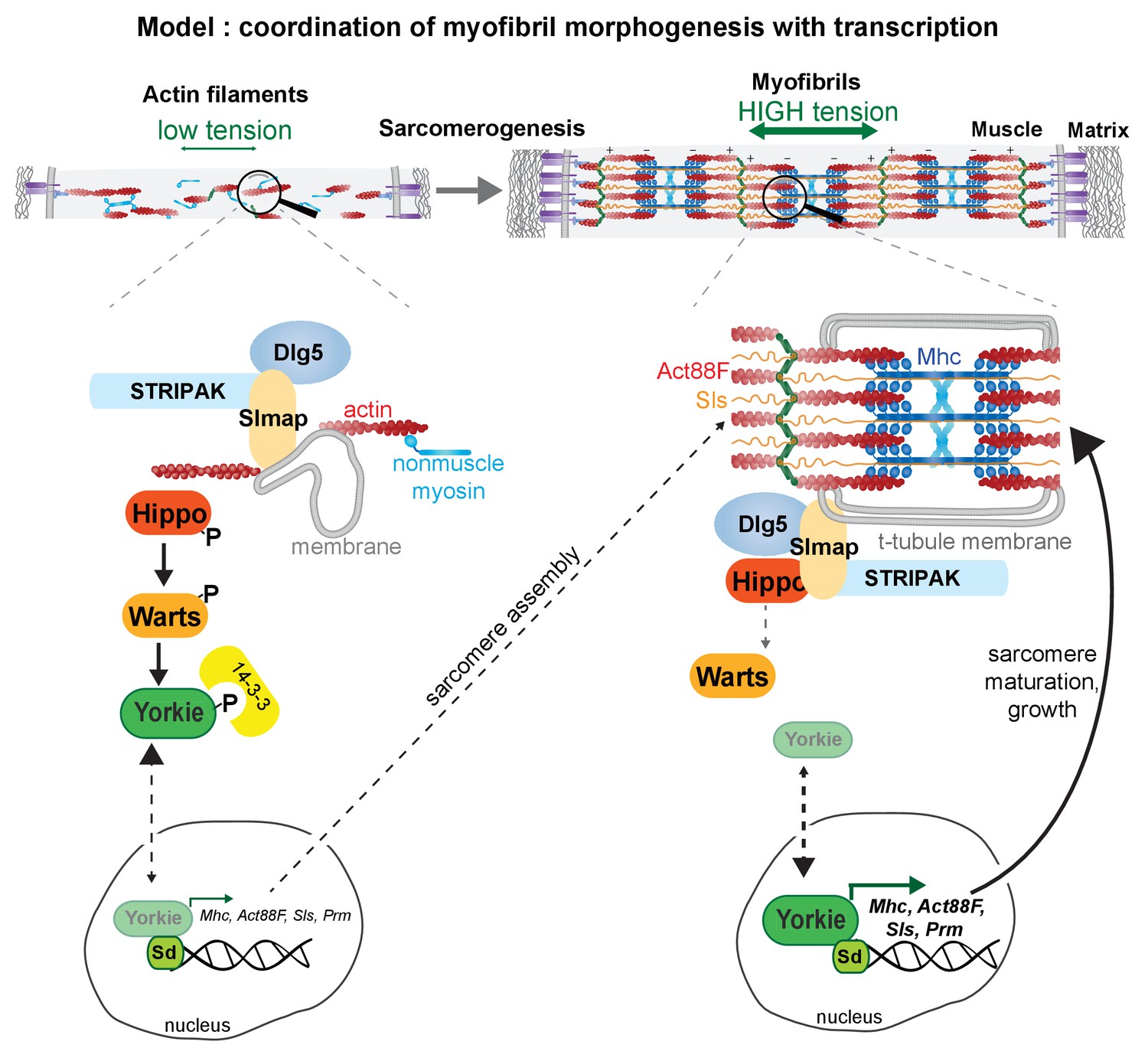
The Hippo Pathway Controls Myofibril Assembly And Muscle Fiber Growth By Regulating Sarcomeric Gene Expression Elife
0 Response to "38 the diagram illustrates a small portion of several myofibrils"
Post a Comment