35 gram negative bacteria diagram
Gram-negative cell walls are strong enough to withstand ∼3 atm of turgor pressure (), tough enough to endure extreme temperatures and pHs (e.g., Thiobacillus ferrooxidans grows at a pH of ≈1.5) and elastic enough to be capable of expanding several times their normal surface area ().Strong, tough, and elastic … the gram-negative cell wall is a remarkable structure which protects the ... Gram-negative infections include those caused by Klebsiella, Acinetobacter, Pseudomonas aeruginosa, and E. coli., as well as many other less common bacteria. Outbreak investigations In the past 3 years, the Division of Healthcare Quality Promotion has assisted in at least 10 investigations of outbreaks of gram negative infections.
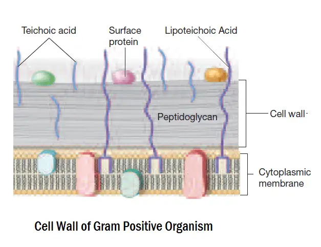
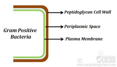
THE GRAM-POSITIVE CELL ENVELOPE · by TJ Silhavy · 2010 · Cited by 2564 — Gram-positive bacteria lack an outer membrane but are surrounded by layers of peptidoglycan many times thicker than ...Abstract · THE GRAM-NEGATIVE CELL... · THE GRAM-POSITIVE CELL...
Gram negative bacteria diagram
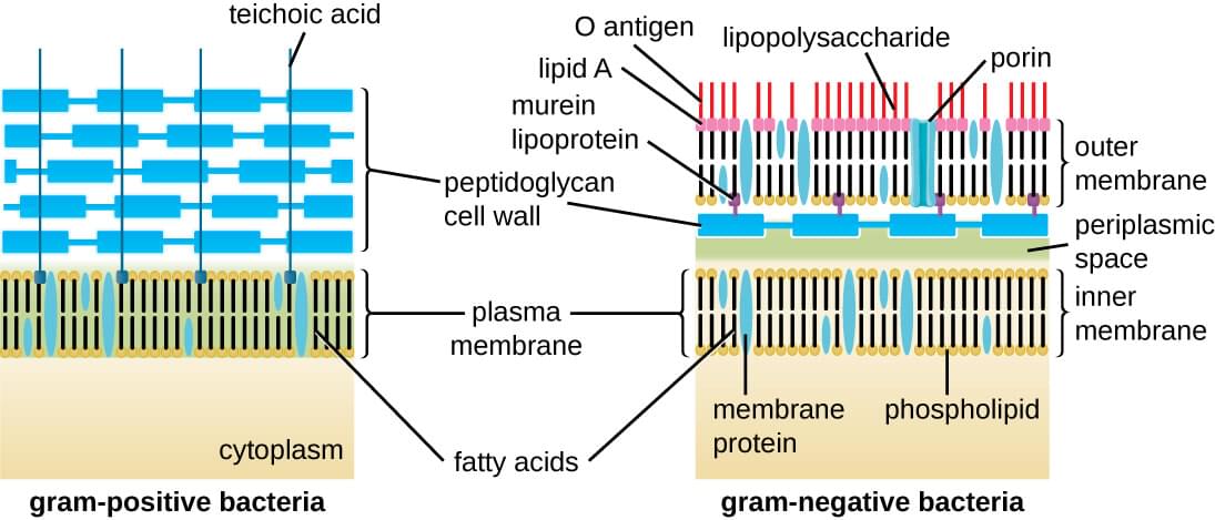
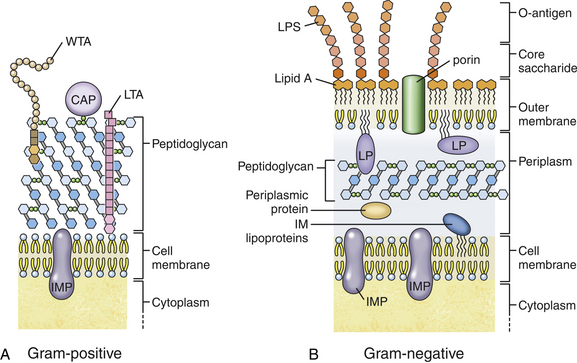
by KC Huang · 2008 · Cited by 291 — In both Gram-negative and Gram-positive bacteria, the cell wall is constructed from the polymer peptidoglycan, a composite of long strands ... Video made by: Alex Wargo Compare and contrast the cell walls of typical Gram-positive and Gram-negative bacteria. 3. Relate bacterial cell wall structure to the Gram-staining reaction. 37 . 38 Bacterial Cell Wall • Peptidoglycan (murein) –rigid structure that lies just outside the cell plasma membrane
Gram negative bacteria diagram. Download scientific diagram | Diagrammatic representation of the gram-negative bacterial cell envelope. The asymmetric outer membrane contains the ... Gram stain and bacterial morphology: Of all the different classification systems, the Gram stain has withstood the test of time. Discovered by H.C. Gram in 1884 it remains an important and useful technique to this day. It allows a large proportion of clinically important bacteria to be classified as either Gram positive or negative based on their ADVERTISEMENTS: In this article we will discuss about the Structure of Bacterial Cell. Bacteria (sing. bacterium) are unicellular prokaryotic microorganisms which divide by binary fission. They do not possess nuclear membrane and the nucleus consists of a single chromosome of circular double-stranded DNA helix (Fig. 1.1). Flagella: ADVERTISEMENTS: These are long filamentous, cytoplasmic ... Experts are tested by Chegg as specialists in their subject area. We review their content and use your feedback to keep the quality high. 100% (4 ratings) Transcribed image text: Identify the Gram-negative and Gram-positive bacteria and their flagellum components on the diagram below. Gram-negative Gram-positive Hook Filament Basal body.
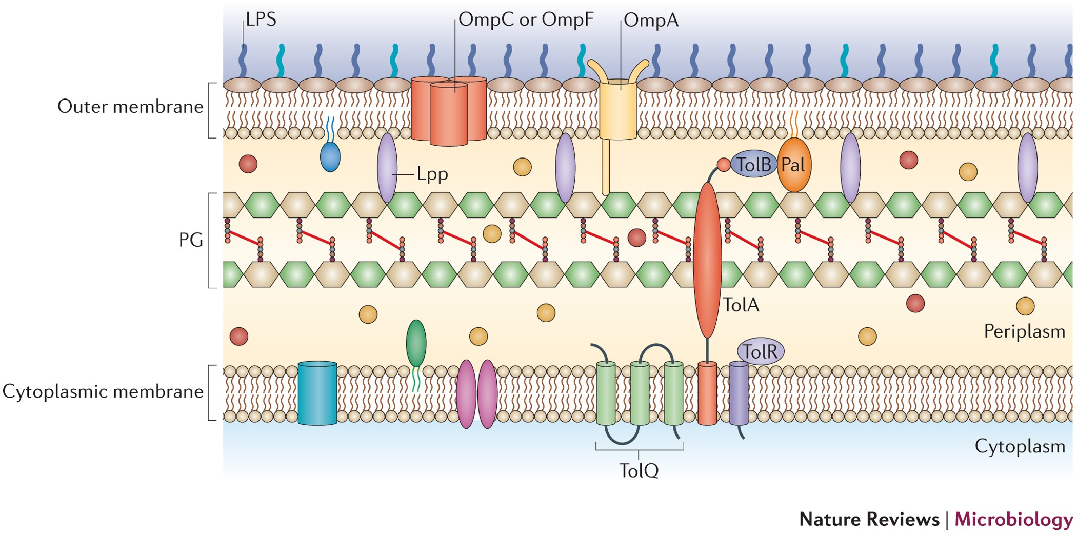
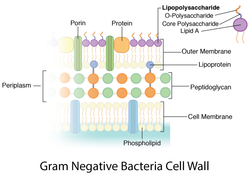
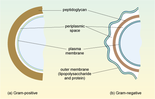
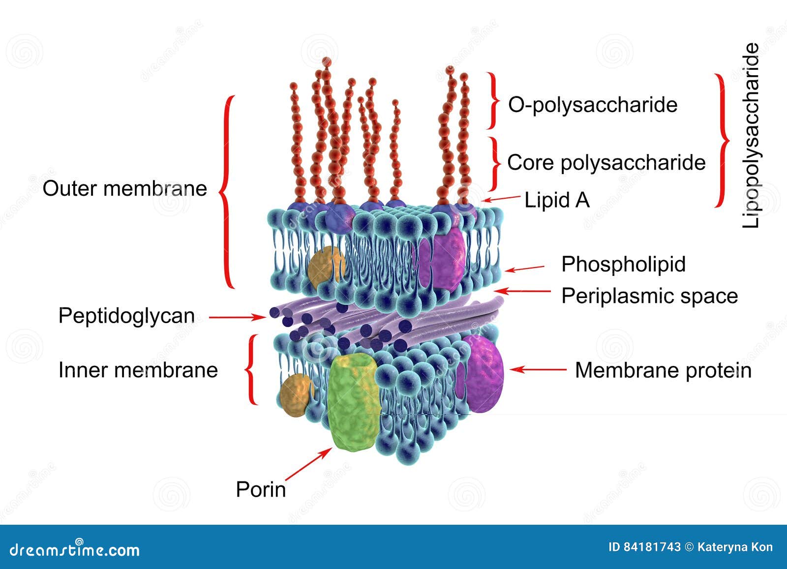
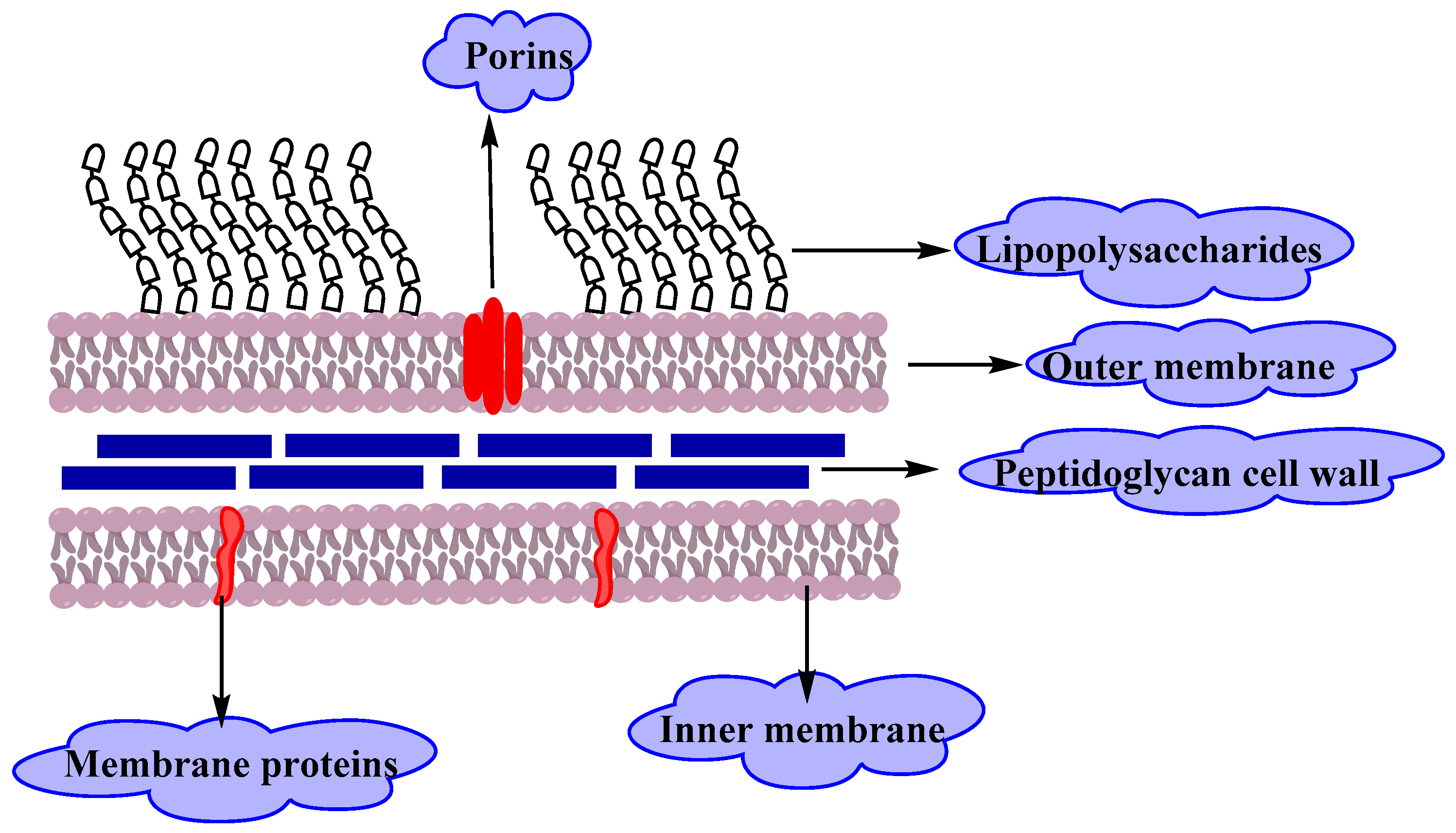
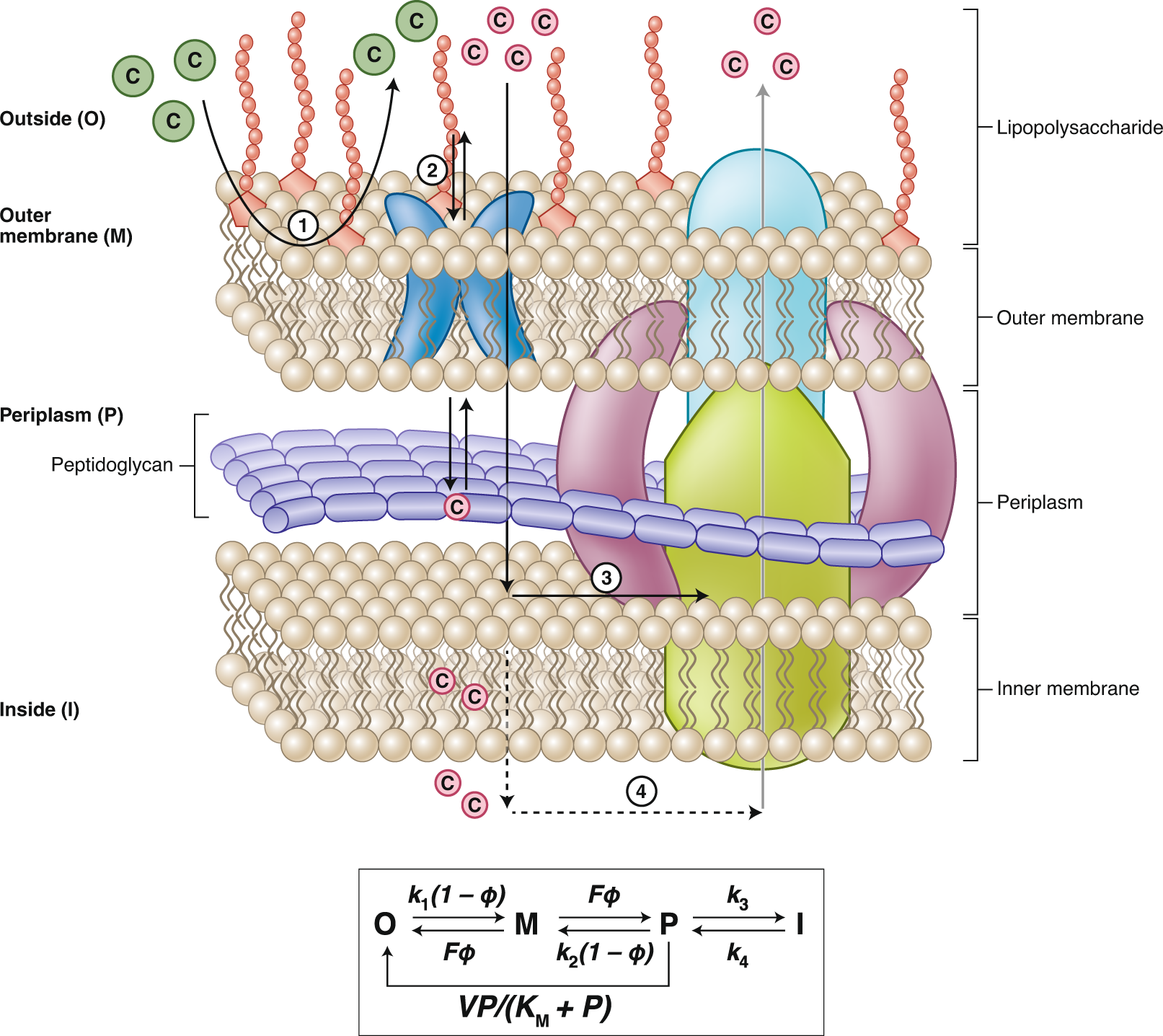
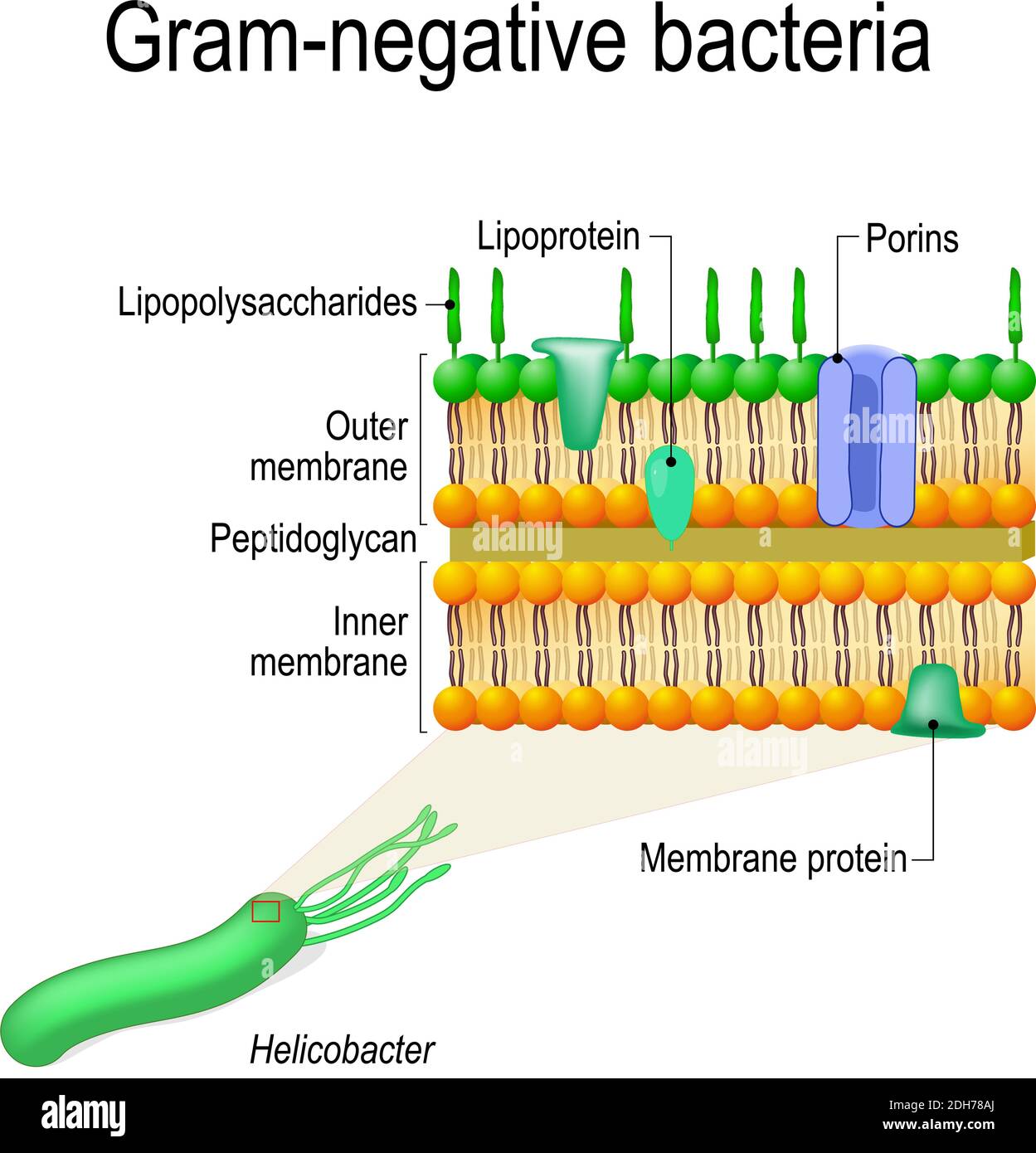
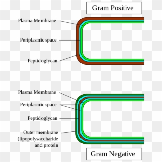
(Ceftriaxone): Good gram negative coverage except pseudomonas, long half-life (q24 hr dosing), crosses blood-brain barrier, biliary and renal clearance. -4. th. generation (Cefipime): Good gram positive (except MRSA) and gram negative coverage, including pseudomonas, crosses blood-brain barrier, good for Gram-negative bacteria have an envelope that consists of three layers (Figure 2).The first layer is the outer membrane (OM), a protective and a unique feature that distinguishes Gram-negative bacteria from Gram-positive bacteria. The OM has phospholipids that are bound to the inner leaflet of the membrane, and lipopolysaccharide (LPS) bound to ... Modelling the structure of the bacterial sacculus · by W Vollmer · 2008 · Cited by 1918 — Gram-positive bacteria contain many surface proteins (e.g. protein A, fibrinonectin-binding ... One of the several unique characteristics of gram-negative bacteria is the structure of the bacterial outer membrane. The outer leaflet of this membrane ...Characteristics · Taxonomy · Bacterial transformation · Role in disease
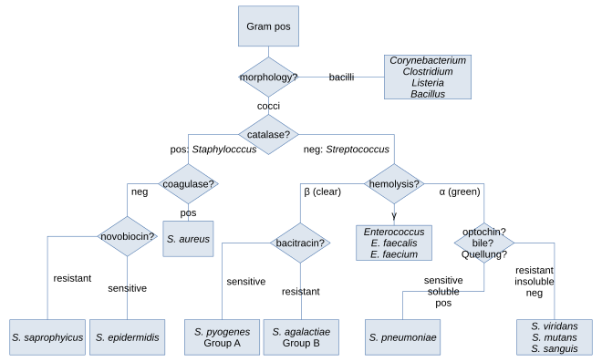
Gram positive bacteria have a thick peptidoglycan layer and no outer lipid membrane whilst Gram negative bacteria have a thin peptidoglycan ...21 Aug 2019 · Uploaded by Beverly Biology Flow Chart Of The Study Design Each Patient Having A First Positive Scientific Diagram. Flowchart For Identification Of Anaerobic Gram Positive Bacilli 1 Scientific Diagram. Gram Negative Bacteria Positive Microbiology Flowchart Others Angle Text Infection Png Pngwing. Starting With Figure 17 5 And Using From Case Study T Chegg. Compare and contrast the cell walls of typical Gram-positive and Gram-negative bacteria. 3. Relate bacterial cell wall structure to the Gram-staining reaction. 37 . 38 Bacterial Cell Wall • Peptidoglycan (murein) –rigid structure that lies just outside the cell plasma membrane Video made by: Alex Wargo
Gram Positive Bacteria Wikipedia
by KC Huang · 2008 · Cited by 291 — In both Gram-negative and Gram-positive bacteria, the cell wall is constructed from the polymer peptidoglycan, a composite of long strands ...

Schematic Structure Of Gram Positive And Gram Negative Cell Walls Download Scientific Diagram

Gram Negative Bacteria Diagram Quizlet
Differences Between Gram Positive And Gram Negative Bacteria Laboratoryinfo Com

Peptidoglycans

Gram Negative Bacteria Wikipedia

Gram Negative Bacteria Microbiology Medbullets Step 1
Bacterial Cell Wall Targets Identification Creative Biolabs
Gram Positive Vs Gram Negative Bacteria A Comparison Easy Biology Class

Difference Bacteria Cell Walls Of Gram Positive And Gram Negative Download Scientific Diagram
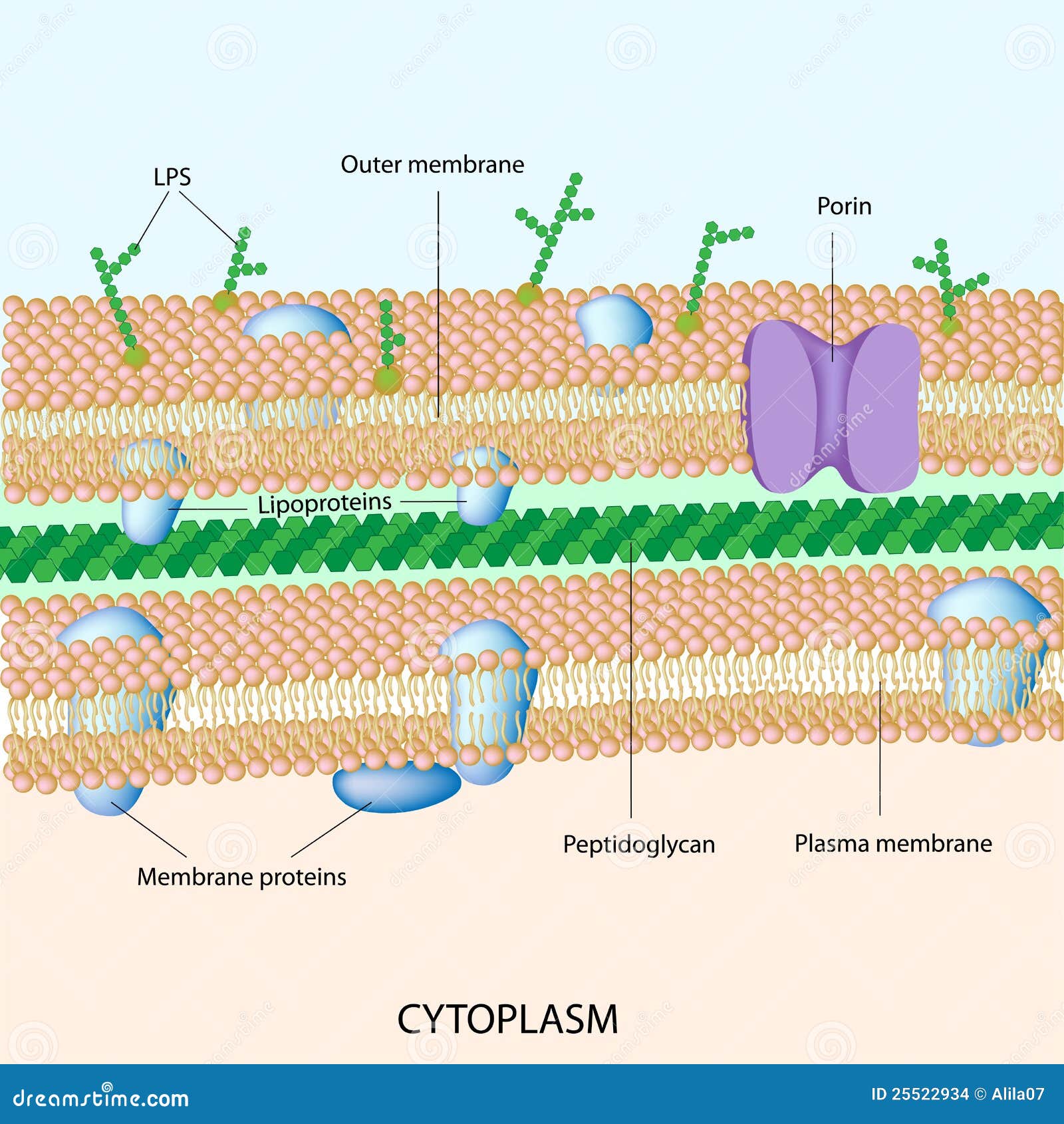
Gram Negative Bacterial Cell Wall Stock Vector Illustration Of Cellular Antibiotics 25522934
1
Outer Membrane Vesicles From Gram Negative Bacteria Biogenesis And Functions Nature Reviews Microbiology
Gram Negative Bacterial Infections Veterian Key
Bacteria Cell Walls General Microbiology

Gram Negative And Gram Positive Bacteria List Of Frontiers Open Access Articles

Structure Of The Cell Wall Of A Gram Negative Bacterium Stock Vector Image Art Alamy
How Can We Change Gram Negative To Gram Positive In Bacteria Quora
Understanding Antibiotic Resistance 4 1 Gram Positive And Gram Negative Bacteria Openlearn Open University Uar 1
Structure Of Gram Negative Bacteria Cell Wall Stock Illustration Illustration Of Lipopolysaccharide Plasma 84181743

Gram Negative Vs Gram Positive Bacteria Cell Model Diagram Quizlet

Why Is It More Difficult To Treat Gram Negative Bacteria Medimoon
Gram Positive Vs Gram Negative Biology Dictionary

Diagram Demonstrating Of The Cell Wall Structure Of A Grampositive Download Scientific Diagram

Gram Positive Vs Gram Negative Bacteria Youtube
Molecules Free Full Text Resistance Of Gram Negative Bacteria To Current Antibacterial Agents And Approaches To Resolve It Html
Defining New Chemical Space For Drug Penetration Into Gram Negative Bacteria Nature Chemical Biology

6 Structure Of Gram Positive And Gram Negative Bacteria Source Download Scientific Diagram

Conjugate Sneaks Antibiotic Into Gram Negative Bacteria
Cell Wall Structure Of Gram Negative Bacteria For Example Helicobacter Vector Diagram For Educational Medical Biological And Science Use Stock Vector Image Art Alamy

Cytosolic Intermediates For Cell Wall Biosynthesis And Degradation Control Inducible B Lactam Resistance In Gram Negative Bacteria Cell
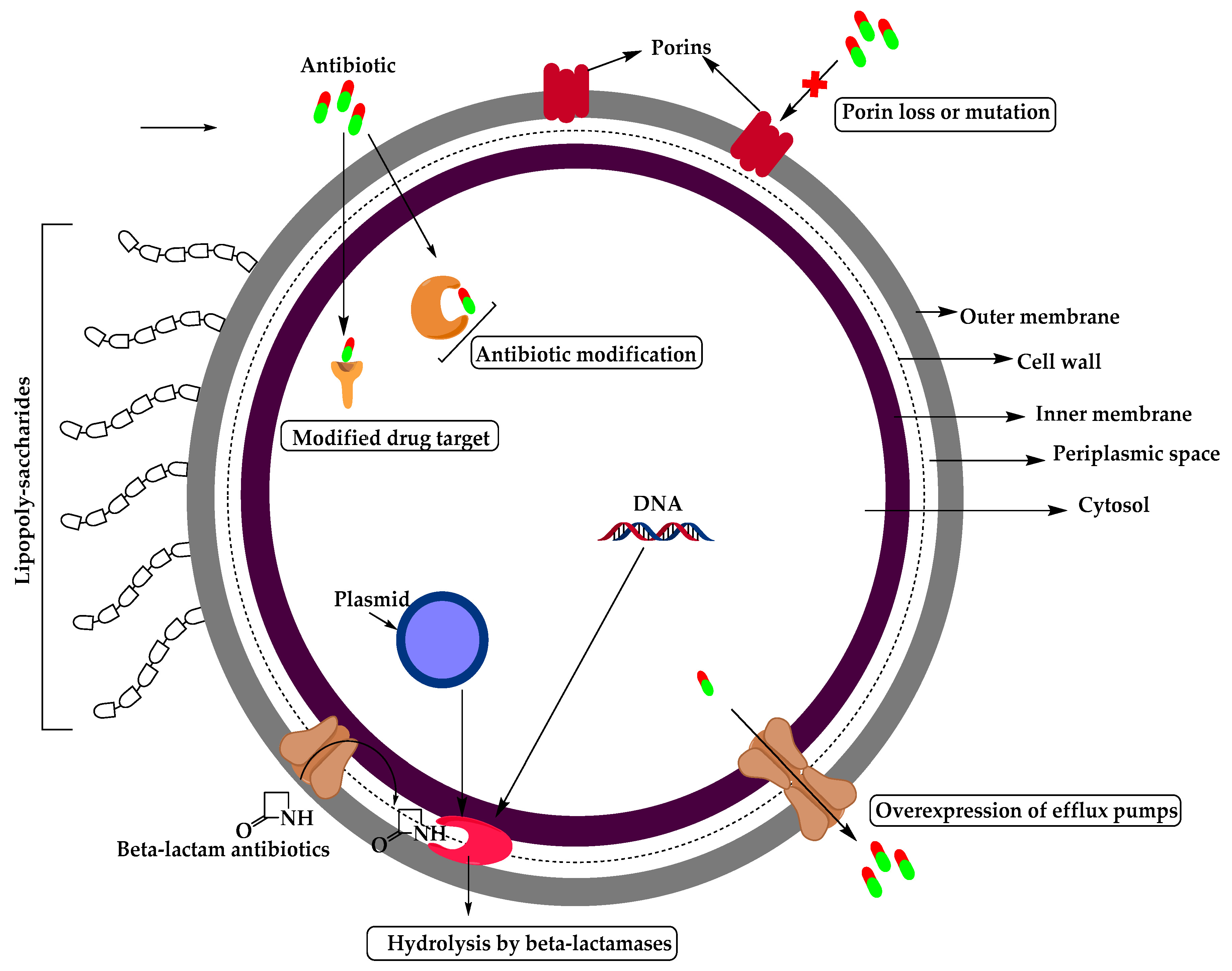
Molecules Free Full Text Resistance Of Gram Negative Bacteria To Current Antibacterial Agents And Approaches To Resolve It Html
Vektor Stok Cell Wall Structure Gramnegative Bacteria Example Tanpa Royalti 1173538834

File Gram Negative Bacteria Lab Methods Algorithm Svg Wikimedia Commons
Gram Cell Wall Gram Positive And Negative Bacteria Hd Png Download 824x1024 4825648 Pngfind
0 Response to "35 gram negative bacteria diagram"
Post a Comment