39 compound microscope ray diagram
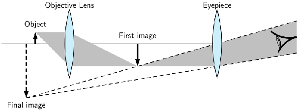
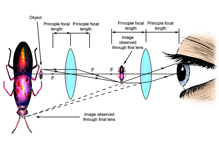
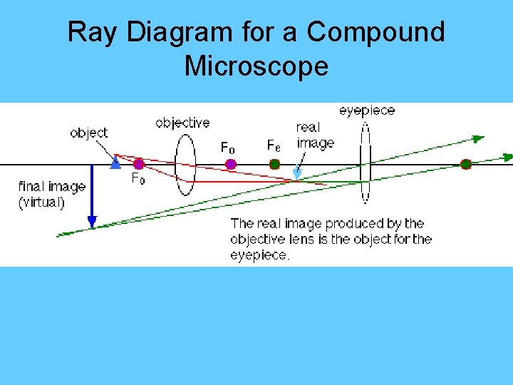
The optical microscope often referred to as the light microscope, is a type of microscope that uses visible light and a system of lenses to magnify images of small subjects. There are two basic types of optical microscopes: Simple microscopes. Compound microscopes. The term "compound" in compound microscopes refers to the microscope having ... Here is the ray diagram of a compound microscope. So, when we are focussing, we move the objective lens which tweaks the image distance. My doubt is that, shouldn't the image be seen clearly, wheresoever the first real image forms, if within Fe (Focus of the eyepiece lens).
(a) Draw a labelled ray diagram of compound microscope, when final image forms at the least distance of distinct vision. (b) Why is its objective of short focal length and of short aperture, compared to its eyepiece? Explain. (c) The focal length of the objective is 4 cm while that of eyepiece is 10 cm. The object is placed at a distance of 6 cm from the objective lens.
Compound microscope ray diagram
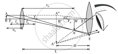
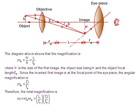
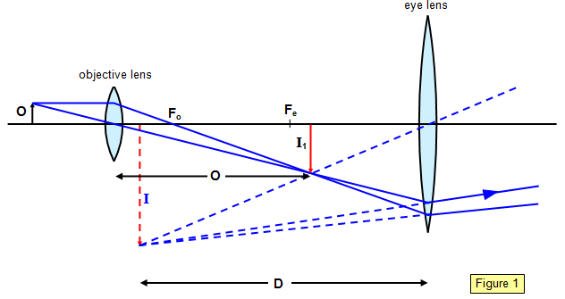
Ray diagram for a compound microscope Total angular magnification, `m = beta/alpha` β → Angle subtended by the image α → Angle subtended by the object Since α and β are small, `tan alpha ≈ alpha and tan beta ≈ beta` `m= (tanbeta)/ (tanalpha)` `tan alpha = (AB)/D` And `tan beta = (A''B'')/D` Ray diagram of a compound microscope. When the final image is formed at the least distance of distinct vision, For the image formed at infinity, u e = f e and By making focal length of the objective small, the magnifying power can be increased. Simple ray diagrams are sufficient to explain many important aspects of microscopy including refraction, focal length, magnification, image formation, and diaphragms. In other instances, it is convenient to refer to light waves as being composed of discrete particles ( quanta ), especially when light is generated by quantum mechanical events or transformed into other forms of energy.
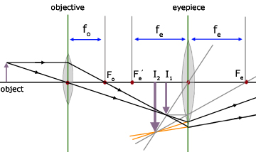
Compound microscope ray diagram. distance or image size. Thus, a ray diagram is used to find the final image distance and to determine the total magnification. The overall magnification of a compound microscope is the product of the individual magnifications of each lens: m = mome (1) where the magnification of either lens is given by: m = - q/p (2) The magnifying power of the compound microscope is defined as the ratio of the angle subtended at the eye by the final image to the angle subtended at the eye by the object when both the final image and the object are situated at the least distance of distinct vision from the eye. Ray diagram for the formation of image by a compound microscope The most familiar type of microscope is the optical, or light, microscope, in which glass lenses are used to form the image. Optical microscopes can be simple, consisting of a single lens, or compound, consisting of several optical components in line. The hand magnifying glass can magnify about 3 to 20×. Single-lensed simple microscopes can ... Nov 18, 2021 · Parts of Atomic Force Microscope. Atomic Force Microscopes have several techniques for measuring force interactions such as van der Waals, thermal, electrical and magnetic force interactions for these interactions done by the AFM, it has the following parts that assist in controlling its functions.
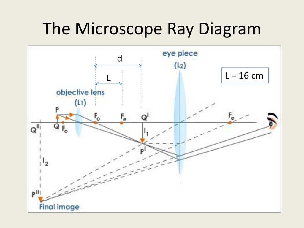
• In compound microscope it will be i.e 10 X, f= 16 mm; 40 X, f= 4 mm; 100 X, f= 1.8 mm. • Image produced by objective lens falls on the eyepiece lens serve as objec t. • Image formed in the ... An electron microscope is a microscope that uses a beam of accelerated electrons as a source of illumination. As the wavelength of an electron can be up to 100,000 times shorter than that of visible light photons, electron microscopes have a higher resolving power than light microscopes and can reveal the structure of smaller objects. Magnification due to a compound microscope. The ray diagram shows that the (linear) magnification due to the objective, namely h'/h, equals Here h' is the size of the first image, the object size being h and f 0 being the focal length of the objective. The first image is formed near the focal point of the eyepiece. Class 12 Physics Ray Optics Optical Instruments. Microscope. Microscope. Microscope is an instrument that gives an enlarged image of minute object. There are 2 types of microscope:-. Simple. Compound. Simple Microscope. An instrument that gives an enlarged image of a minute object.
Simple Microscope. Compound Microscope. 1. Simple Microscope comprises a single biconvex lens used as a magnifying glass. Compound Microscope comprises 2 or more convex lenses where one lens is an eyepiece and the other one is the objective lens. 2. Natural light is the source to see the object. An illuminator is a source to see the object. 3. Here are two ray diagrams for compound microscope, the first one proposed by the book, and the second one recommended by the teacher: In the first image, the light rays form a real image A'B', which becomes the virtual object for the eyepiece. See, the original rays are carried forward to the eyepiece, which then form a virtual image, A"B". A compound microscope is a high magnifying microscope that is used to examine the smaller samples that are not visible to the naked eye such as diatoms, bacteria, or viruses. In this microscope, the sample is kept in a glass slide and is then kept on the stage of the microscope to examine through the lenses. Early history. In ancient India, the philosophical schools of Samkhya and Vaisheshika, from around the 6th–5th century BC, developed theories on light.According to the Samkhya school, light is one of the five fundamental "subtle" elements (tanmatra) out of which emerge the gross elements.
Exercises, Telescopes and microscopes, By OpenStax (Page 2/2 ...
The ray diagram to show the working of compound microscope is shown in figure. A tiny object AB to be magnified is placed in front of the objective lens just beyond its principal focus fo'. In this case, the objective lens O of the compound microscope forms a real, inverted and enlarged image A'B' of the object.

Ray Diagram Of Compound Microscope Pdf - Juanribon.com ...
A ray diagram like this can be interpreted in both directions. If you put a point light source one focal length from a converging lens, parallel light will emerge from the other side. The two sides of a lens are ... The image you see in a compound microscope is a virtual image. Figure 4.12
Draw a Ray Diagram to Show the Working of a Compound ...
Draw a ray diagram of compound microscope, when the final image is formed at the minimum distance of distinct vision. Hint: A compound microscope is an optical instrument used for observing highly magnified images of tiny objects. The compound microscope has two lenses and it works in such a way that the image of the object from the first lens ...

History of Imaging – Part 2
A simple microscope has a limited maximum magnification (≤ 9) for realistic focal lengths. For much larger magnifications, one uses two lenses, one compounding the effect of the other. This is known as a compound microscope. A schematic diagram of a compound microscope is shown in Fig.
a) Draw a ray diagram showing the image formation by a ...
Best answer. (a) Labelled diagram of compound microscope. The objective lens form image A' B' near the first focal point ofeyepiece. (b) Angular magnification of objective lens m0 = linear magnification h'/h. where L is the distance between second focal point of the objective and first focal point of eyepiece.

KOPAL Classes - Compound Microscope Ray Diagram | Facebook
In the following ray diagrams of image formation by a compound microscope, only two main rays are shown. Which ray diagram (s) is (are) correct? Case A Case B Case C Case A and Case B Case A and Case C Case Band Case C In a compound microscope, what would happen If the focal length of the eyepiece is increased? The image would be enlarged The ...

Ray Diagram of a Compound Microscope | Diagram, Microscope ...
Draw a ray diagram of compound microscope, when final image is formed at the minimum distance of distinct vision. Easy Solution Verified by Toppr It consist of two convex lenses, one objective of very small focal length with short aperture. And one Eyepiece with moderate focal length and large aperture. Was this answer helpful? 0 0

Optical Instruments: Compound Microscope and its Magnification
(a) Draw a ray diagram of a compound microscope for the final image formed at least distance of distinct vision? Answer Diagram of Compound Microscope for the final image formed at D: (b) An angular magnification of 30X is desired using an objective of focal length 1.25 cm and an eye piece of focal length 5 cm.
Draw a neat labelled diagram of a compound microscope and ...
Draw a neat labeled ray diagram of a compound microscope. Explain briefly its working. asked Aug 1, 2019 in Physics by Rk Roy (63.7k points) jee; jee mains; 0 votes. 1 answer (i) Draw a neat labelled ray diagram of an astronomial telescope in normal adjustment . Explain briefly its working . (ii) An astronomical telescope u
The Compound Microscope - ppt download
Transmission electron microscopy (TEM) analysis is conducted to get the actual size of the nanocrystalline cellulose fibers and in some cases the morphology. Nasseri and Mohammadi [99] obtained individual cellulose whiskers with length (L) of 87±28 nm and diameter (d) of 15±3 nm, with an average aspect ratio (L/d) of whiskers obtained was 6±2.

Ray diagram of focussing on a compound microscope - Physics ...
This Fastest drawing Technique to draw Compound Microscope Ray Diagram is for all students of every Board - WBCHSE, HSC, CBSC, ICSE and other state boards fr...

Draw the ray diagram of image formation in case of compound microscope.
Draw a ray diagram to show the image formation by a compound microscope when the final image is formed at the near point.Define the resolving power of a microscope.Write two factors by which resolving power can be increased?
Compound Microscope, Ray Diagram Mistakes. | Physics Forums
In a microscope, light is focused on the object as a narrow pencil of light, from where it enters into the objective as a diverging pencil (Figure 4.8). The angle 9 subtended by the optical axis (the line joining the centers of all the lenses) and the outermost ray still covered by the objective is a measure of the aperture called 'half ...

Download HD Draw A Ray Diagram Of A Compound Microscope ...
How to draw ray diagram of Compound Microscope - Chapter 7 - Lenses - Part 4
Convex lens use - Microscope
Topic compound microscope explains detail working with ray diagram and magnification of compound microscope. Helpful for cbse class 12 physics chapter 9 ray optics. CBSE 12 Physics 01 Electric Charges and Fields 17 Topics 01.01 Electric Charge. 01.02 Conductors, Semiconductors and Insulators.

Draw a labelled diagram of an image formed by a compound ...
Nov 26, 2021 · Observation under the compound microscope. Different structures within the pollen appear better under staining as it provides contrast. Under a compound microscope, pollen appears ovoid and is provided on the surface with scales or similar structures. The structure of the pollen also depends on the type of plant.
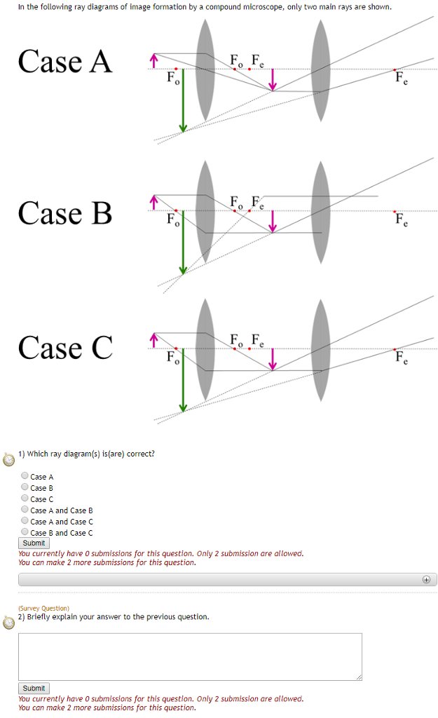
Solved In the following ray diagrams of image formation by a ...
Compound microscope is a type of optical microscope that is used for obtaining a high-resolution image. There are more than two lenses in a compound microscope. Learn about the working principle, parts and uses of a compound microscope along with a labeled diagram here.

Draw a schematic ray diagram of a compound microscope with ...
Simple ray diagrams are sufficient to explain many important aspects of microscopy including refraction, focal length, magnification, image formation, and diaphragms. In other instances, it is convenient to refer to light waves as being composed of discrete particles ( quanta ), especially when light is generated by quantum mechanical events or transformed into other forms of energy.
a) Draw a ray diagram showing the image formation by a ...
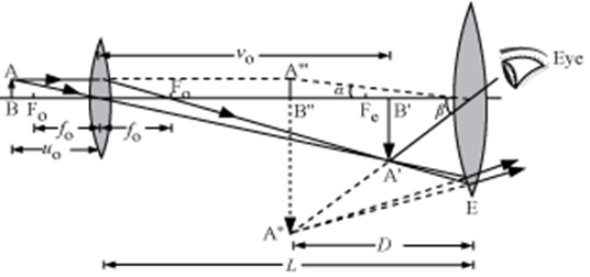
Ray diagram of a compound microscope. When the final image is formed at the least distance of distinct vision, For the image formed at infinity, u e = f e and By making focal length of the objective small, the magnifying power can be increased.

Draw a ray diagram to show formation of an image by a ...
Ray diagram for a compound microscope Total angular magnification, `m = beta/alpha` β → Angle subtended by the image α → Angle subtended by the object Since α and β are small, `tan alpha ≈ alpha and tan beta ≈ beta` `m= (tanbeta)/ (tanalpha)` `tan alpha = (AB)/D` And `tan beta = (A''B'')/D`

Draw a ray diagram of compound microscope, when final image ...
What is Compound microscope ? Draw a ray diagram showing the ...

Ray diagram for compound microscope when the image is formed ...
Electricity - detailed contents

what ray diagram indicates of compound microscope?​ - Brainly.in

Draw a ray diagram of a compound microscope for the final ...

Draw the ray diagram of image formation in case of compound ...
a draw a ray diagram showing the image formation by a ...

Experts, please give ray diagram of compound microscope ...

Draw a labelled ray diagram to show image formation by a ...

Show by Ray diagram, the formation of image in case of a ...

draw the labelled ray diagram for the formation of image by a ...
Q. 18.(a) Draw a labelled ray diagram to show the formation ...

Draw a neat labelled diagram of a compound microscope and ...

compound microscope image at infinity reply fast with diagram ...

9.4 Microscopes | Texas Gateway
Optical Instruments Power of a lens Optometrists instead

Draw a Ray Diagram Showing Image Formation in a Compound ...
schoolphysics ::Welcome::
0 Response to "39 compound microscope ray diagram"
Post a Comment