39 gel electrophoresis labeled diagram
Gel electrophoresis labeled diagram. An enzyme is used to separate a strand of dna from a source and the dna is suspended in a dye. A lot of expertise and experience are required for interpreting gel electrophoresis. Electrophoresis units are available for running either vertical or horizontal gel system. 4. You will now simulate gel electrophoresis. Locate the simulated electrophoresis gel with all the suspects labeled on your summary sheet. The numbers along the sides of YOUR gel indicate lengths of fragments (number of bases in each fragment). Plot your fragment lengths for each sample by drawing lines in the appropriate region of your gel.
Place gel into electrophoresis unit. Add 150 ml 1X TBE buffer to completely fill the box and to cover the top gel surface with about 2 mm of buffer. Note: At this point the gel box can be covered and left until the next day if necessary. On the gel load 5-10 µl of each dye into a well.
Gel electrophoresis labeled diagram
Based on the data they collected using gel electrophoresis, label the branching tree diagram below. Write the letters A, B, and C, to represent the possible evolutionary relationships between species A, B, and C Gel Electrophoresis Steps. The broad steps involved in a common DNA gel electrophoresis protocol: 1. Preparing the samples for running. The DNA is isolated and preprocessed (e.g. PCR, enzymatic digestion) and made up in solution with some basic blue dye to help visualize the movement of the sample through the gel. 2. When gel has cooled, place it into the electrophoresis cell and remove the comb. 8. Add about 325-350mls of 1X sodium borate buffer to the electrophoresis chamber, buffer should just cover the gel. 9. Label five 0.5ml tubes, one for each of your PCR amplifications. 10.
Gel electrophoresis labeled diagram. Table 1. Results Of Gel Electrophoresis Of Example 4 kb Plasmid Enzyme EcoR I + BamH I R I + Hind III H I + Hind III Fragments Produced (bp) *2,000 5001,500 3,500 2,500 1,500 2,000 *Indicates these two cutting sites are equal distance apart on the plasmid so that what would actually be two bands appears as a single band on the gel. Start studying Gel Electrophoresis. Learn vocabulary, terms, and more with flashcards, games, and other study tools. Agarose Gel Electrophoresis. Agarose gel electrophoresis separates DNA fragments according to their size. Typically, a DNA molecule is digested with restriction enzymes, and the agarose gel electrophoresis is used as a diagnostic tool to visualize the fragments.An electric current is used to move the DNA molecules across an agarose gel, which is a polysaccharide matrix that functions as a sort ... DNA profiling is a technique used to identify individuals by the characteristics of their DNA.PCR (Polymerase Chain Reaction) is a method to make multiple copies of DNA from a single copy or a few copies. Scientist use gel electrophoresis whenever they need to sort DNA strands according to length.These methods would be used when there is an unknown source of DNA such as blood at a crime scene.
5. More restriction digestion and gel electrophoresis; follow up to diagram 5. *There are three similar diagrams here. Your test will have one of these three gel images. Gel A You decide to follow up your first experiment (results from good experiments should be reproducible). You obtain a sample of linear DNA with the following sequence. Draw a neat labelled diagram of a typical agarose gel electrophoresis. Easy. Answer. In agarose gel electrophoresis, agarose is used as a matrix. The sample is added in the slot and current is applied to it. The smaller molecules move faster and the larger molecules are retarded. In this method, separation is based on charge and size of the ... (a) Using the circle provided, construct a labeled diagram of the restriction map of the plasmid. Explain how you developed your map. Construct a labeled map and explain (3 points maximum) H H E E 40 20 30 10 H E 40 20 30 10 E H E = EcoRI Restriction Point H = HaeIII Restriction Point Written By Pelvic Diagram Saturday, September 5, 2020. Edit. Gel Electrophoresis Diagram. Gel electrophoresis is a technique used to separate mixtures like DNA and proteins. Gel electrophoresis uses a gel (like gelatin) and an electric field is put through the gel.
Gel Electrophoresis. Lane 1: DNA Ladder. Lane 2: Undigested plasmid A. Lane 3: Completely digested plasmid A. Lane 4: Digested PCR product (or DNA Fragment). Lane 5: PCR Product (with a faint primer dimer band). Lane 6: Genomic DNA. The white arrows indicate the bands that you want to excise. Jul 21, 2021 — Gel electrophoresis is a technique commonly used in laboratories to separate charged molecules like DNA?, RNA? and proteins? according to ... Agarose gel electrophoresis is the most effective way of separating DNA fragments of varying sizes ranging from 100 bp to 25 kb 1.Agarose is isolated from the seaweed genera Gelidium and Gracilaria, and consists of repeated agarobiose (L- and D-galactose) subunits 2.During gelation, agarose polymers associate non-covalently and form a network of bundles whose pore sizes determine a gel's ... a) Each band in a DNA electrophoresis gel is made up of one molecule of DNA. b) Gel electrophoresis can tell you the sequence of a particular DNA fragment. c) ...2 pages
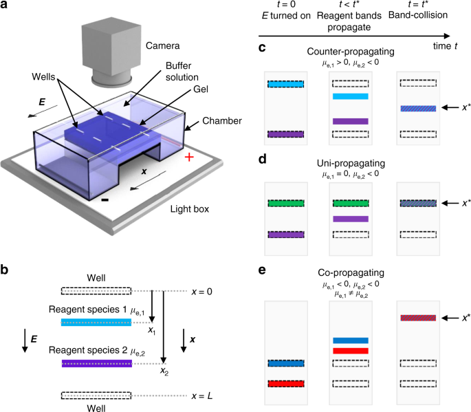
Band Collision Gel Electrophoresis Nature Communications
Electrophoresis (With Diagram) The term electrophoresis describes the migration of a charged particle under the influence of an electrical field. Many important biomolecules — such as peptide, proteins nucleotides and nucleic acids — possess ionisable groups and, therefore, at any given pH, exist in solution as electrically charged species ...
Southern Blot Analysis Springerlink
4. Use the shapes/ text box features in Word to draw and label a diagram of a gel electrophoresis unit. Make sure to include: a. The wells (1 pt.) b. The anode and cathode (and their charge) (1 pt.) A ladder with fragments at 10, 50, 100, and 500 base pairs (bp) (1 pt.) d. Sample 1: with fragments at 75 bp and 75 bp (1 pt.) e.

Electrophoresis And Blotting Of Dna Tamber Major Reference Works Wiley Online Library
• The process of gel electrophoresis, including DNA ... of amplified sample in the new labeled tubes. ⎕ STEP 7 Label the diagram on the last page of this ... Use a separate well for the DNA ladder and negative control. c. Label your gel (to differentiate from the gel of classmates). You can write on the orange frame of LONZA gel with a ...
12 2 Visualizing And Characterizing Dna Biology Libretexts
Gel electrophoresis is a laboratory procedure used to separate biological molecules with an electrical current. Previously, we've discussed gel electrophoresis in the context of analyzing DNA .
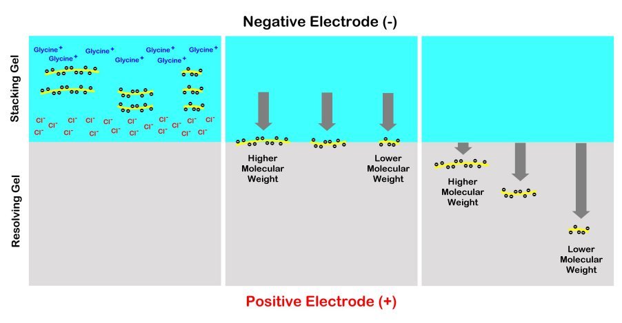
1 15 Sds Page Biology Libretexts
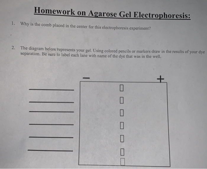
a. After you find out what dyes you are using, draw a picture of the gel and the wells. Label which dyes you will put in each well. b. When you load a gel, it is very important that you do not damage the gel in any way. You must be very careful not to "jab" the gel with the end of your pipet. Ideally, you shouldn't even touch the gel with the ...

What Is Gel Electrophoresis Facts Yourgenome Org
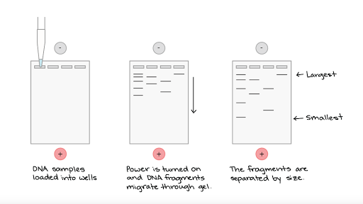
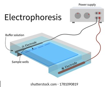
Gel electrophoresis is a technique used to separate DNA fragments according to their size. DNA samples are loaded into wells (indentations) at one end of a gel, and an electric current is applied to pull them through the gel. DNA fragments are negatively charged, so they move towards the positive electrode. Because all DNA fragments have the ...

Confirmation Of Qd Labeling Of Pdna By Agarose Gel Electrophoresis Qd Download Scientific Diagram
Diagram of agarose gel setup, for agarose gel electrophoresis. (Figure by MIT OpenCourseWare.) Today you will separate DNA fragments using an agarose matrix. Agarose is a polymer that comes from seaweed and if you've ever made Jell-O™, then you already have all the skills for pouring an agarose gel.
Gel Electrophoresis
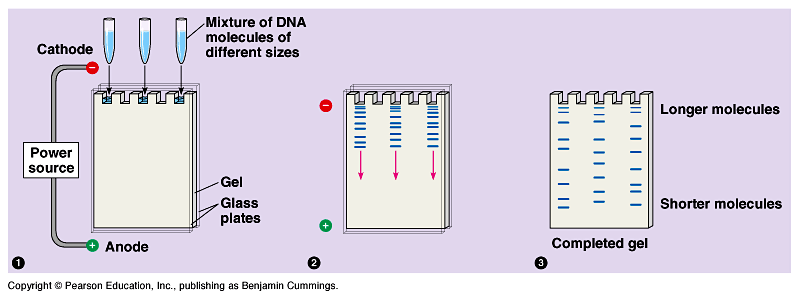
Gel electrophoresis is a type of biotechnology that separates molecules based on their size to interpret an organism's DNA. An enzyme is used to separate a strand of DNA from a source and the DNA is suspended in a dye. Then, the dye is applied to a negatively-charged gel on one side of a sheet.
Gel Electrophoresis Article Khan Academy
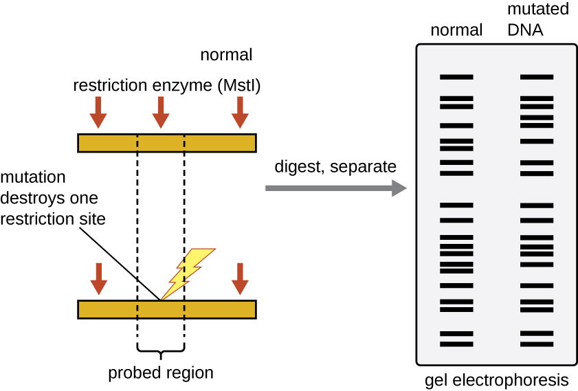
10. On the gel picture below, (a) circle the smallest fragment produced by a restriction enzyme and label it "smallest." (b) circle the largest fragment produced by a restriction enzyme and label it "largest." 11. In one or two sentences, summarize the technique of gel electrophoresis. Student answers DNA restriction fragment size chart

Cont Gel Electrophoresis Basic Concept Ppt Video Online Download
Image 2: An agarose gel electrophoresis is a process useful in various applications including forensic investigation, molecular cloning, and genetic fingerprinting. Picture Source: news-medical.net Applications of agarose gel electrophoresis. It helps identify unknown samples. It is a method of choice for checking the quality and accuracy of other procedures.
Schematic Illustration Of The Difference Gel Electrophoresis Dige Download Scientific Diagram
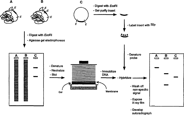
electrophoresis, the radio-labeled fragments are visualized by exposing the gel to an x-ray fil-ter to make an autoradiogram. Figure 3 shows the pattern of bands that would be created on the autoradiogram by the four sets of labeled fragments in Figure 2. Recall that each band contains many copies of one of those labeled fragments.

Addgene Protocol How To Run An Agarose Gel
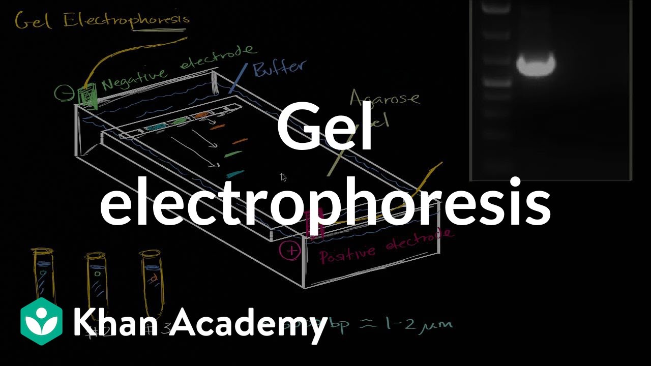
Today, we'll be talking about gel electrophoresis. What is gel electrophoresis, you might ask. Well, it's a lab technique usually used in the biochemistry lab for separating out DNA or proteins based on their size. And let's talk about how it works. So first, you need to have the gel.

Genetic Identification Ofprotein Bands In Gel Electrophoresis Download Scientific Diagram
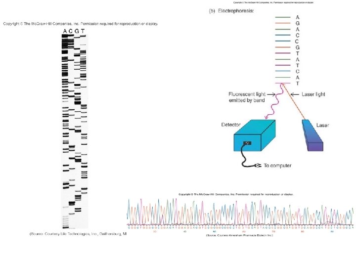
This diagram summarizes the Sanger sequencing method using fluorochrome-labeled ddNTPs and capillary gel electrophoresis. Since 2005, automated sequencing ...

Two Dimensional Gel Electrophoresis An Overview Sciencedirect Topics
About plasmid DNA and gel electrophoresis: . Plasmid DNA can exist in three conformations: supercoiled, open-circular (oc), and linear (supercoiled plasmid DNA is often referred to as covalently closed circular DNA, ccc). In vivo, plasmid DNA is a tightly supercoiled circle to enable it to fit inside the cell. In the laboratory, following a careful plasmid prep, most of the DNA will remain ...

Passivated Gel Electrophoresis Of Charged Nanospheres By Light Scattering Video Tracking Sciencedirect
When gel has cooled, place it into the electrophoresis cell and remove the comb. 8. Add about 325-350mls of 1X sodium borate buffer to the electrophoresis chamber, buffer should just cover the gel. 9. Label five 0.5ml tubes, one for each of your PCR amplifications. 10.

Gel Electrophoresis Video Khan Academy
Gel Electrophoresis Steps. The broad steps involved in a common DNA gel electrophoresis protocol: 1. Preparing the samples for running. The DNA is isolated and preprocessed (e.g. PCR, enzymatic digestion) and made up in solution with some basic blue dye to help visualize the movement of the sample through the gel. 2.

Nucleic Acids And Chromatin 2 4 Analysis Of Nucleic Acids By Electrophoresis And Hybridisation Openlearn Open University S377 1
Based on the data they collected using gel electrophoresis, label the branching tree diagram below. Write the letters A, B, and C, to represent the possible evolutionary relationships between species A, B, and C
Homework On Agarose Gel Electrophoresis 1 Why Is Chegg Com

What Is Gel Electrophoresis Facts Yourgenome Org
Chem Purdue Edu

Use Of Two Dimensional Semi Denaturing Detergent Agarose Gel Electrophoresis To Confirm Size Heterogeneity Of Amyloid Or Amyloid Like Fibers Protocol
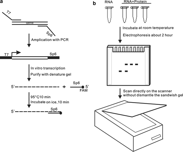
Using Fam Labeled Dna Oligos To Do Rna Electrophoretic Mobility Shift Assay Springerlink

Draw A Diagram Of A Typical Agarose Gel Electrophoresis Showing Migration Of Undigested
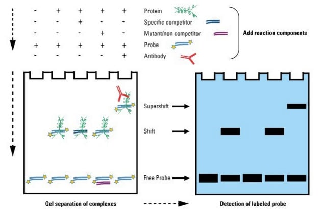
Gel Shift Assays Emsa Thermo Fisher Scientific Fr
Agarose Gel Electrophoresis Age Image Of The Pcr Products A With The Download Scientific Diagram
A A Denaturing Agarose Gel Electrophoresis Displaying 28s And 18s Download Scientific Diagram

How To Interpret Dna Gel Electrophoresis Results Goldbio

Small Scale Agarose Gel Electrophoresis 32p Labeled Ere Containing Download Scientific Diagram
What Kind Of Dna Fragments Could Reach Positive End Of Gel During Electrophoresis Socratic

Gel Electrophoretic Pattern And Northern Blotting Of Dsrna Fragments Download Scientific Diagram
Gel Electrophoresis Video Biotechnology Khan Academy
Pcr Primer Design Attgccacgttgacggtcag Cacgatttccggaagtac Inducer Iptg Lactose

Agar Gel Electrophoresis An Overview Sciencedirect Topics
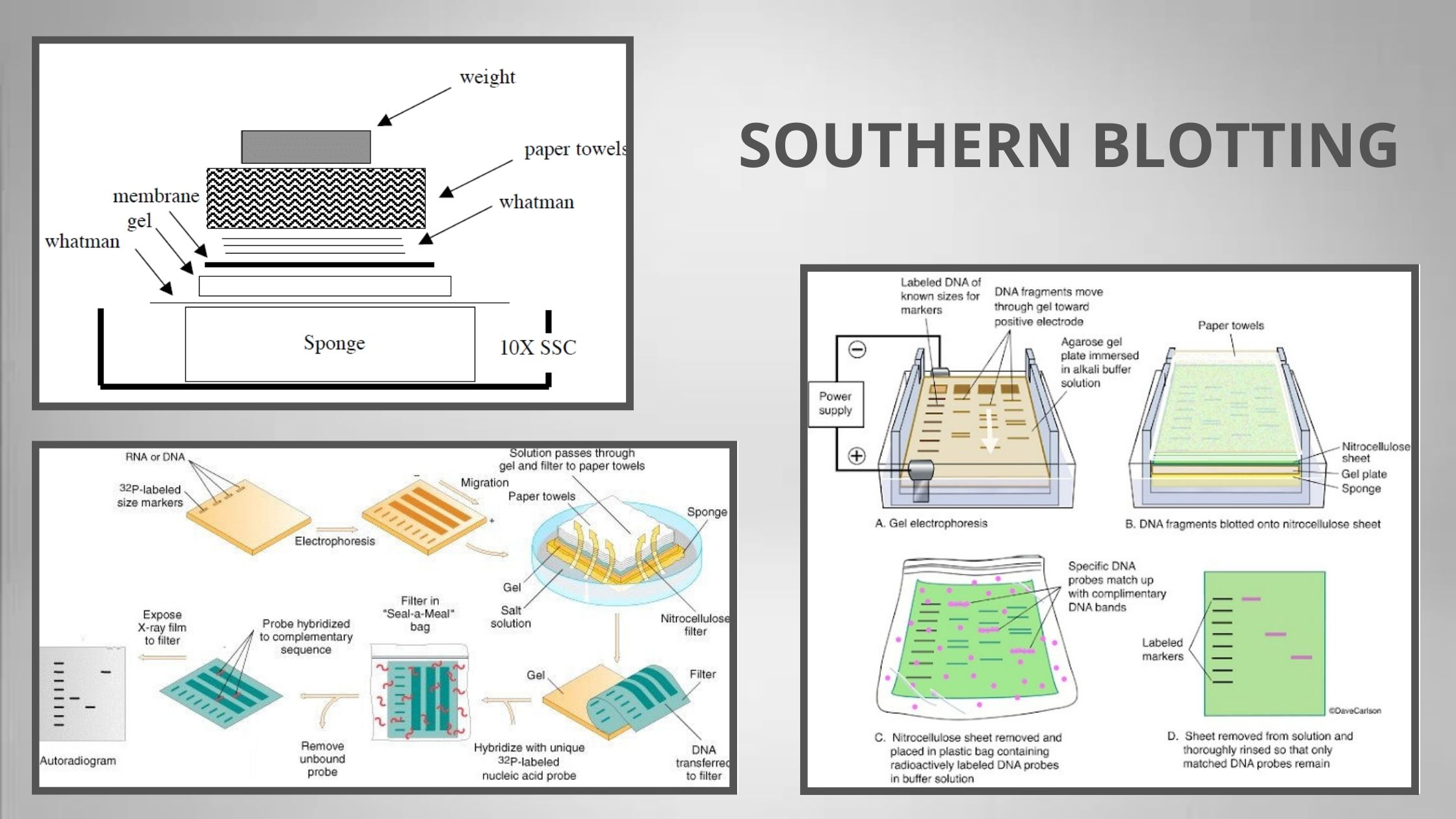
Southern Blotting Technique Principle Procedure Importance

Gel Electrophoresis Bioninja
Electrophoresis Gel Images Stock Photos Vectors Shutterstock

Indesign Labeling Annotating Pcr Gel Pictures Advanced Tutorial Part 12 Youtube

Gel Electrophoresis To Determine Genotype Youtube

Sds Gel Electrophoresis Of 36 Kda Protein Labelled With 32 P In Vivo Or Download Scientific Diagram
0 Response to "39 gel electrophoresis labeled diagram"
Post a Comment