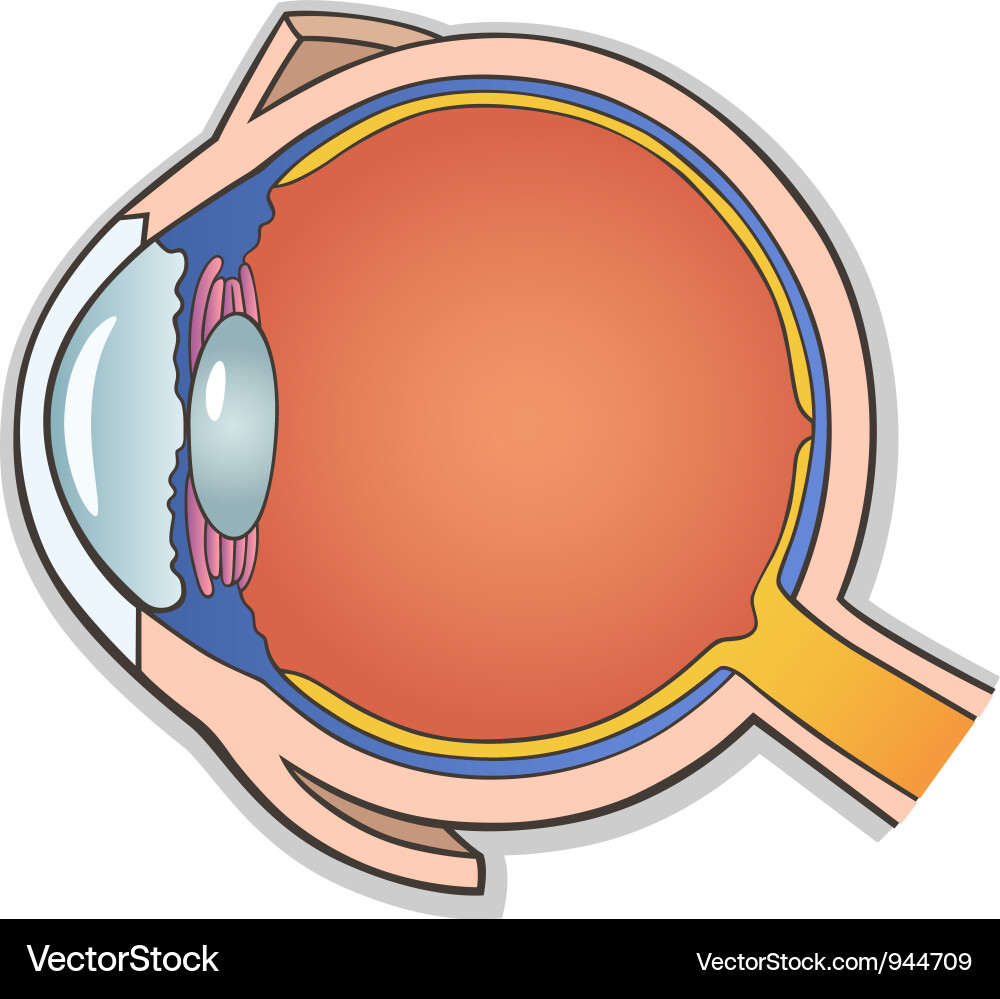
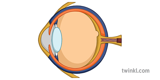
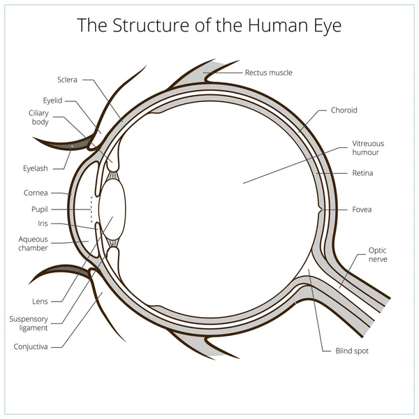
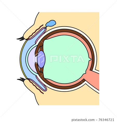
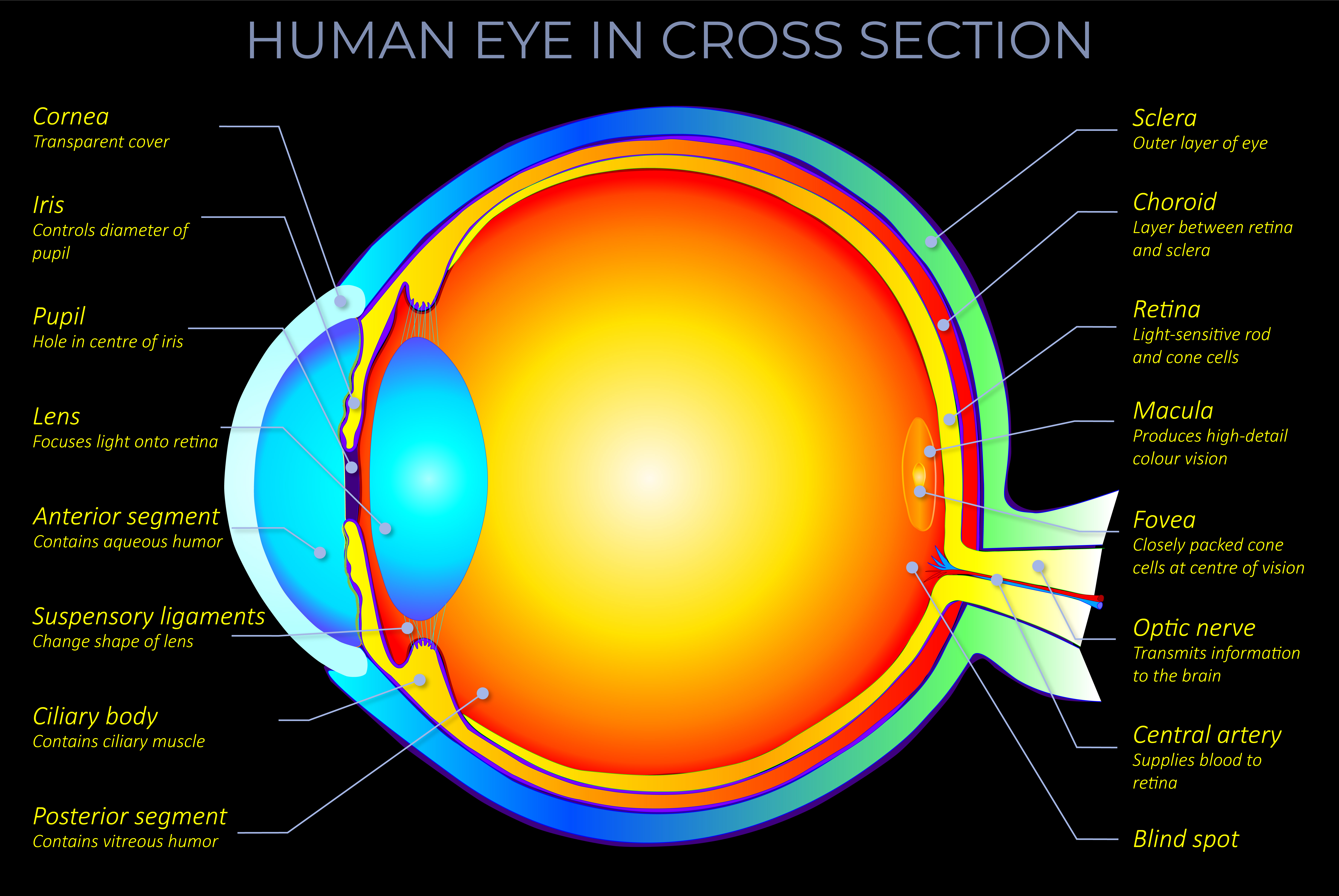
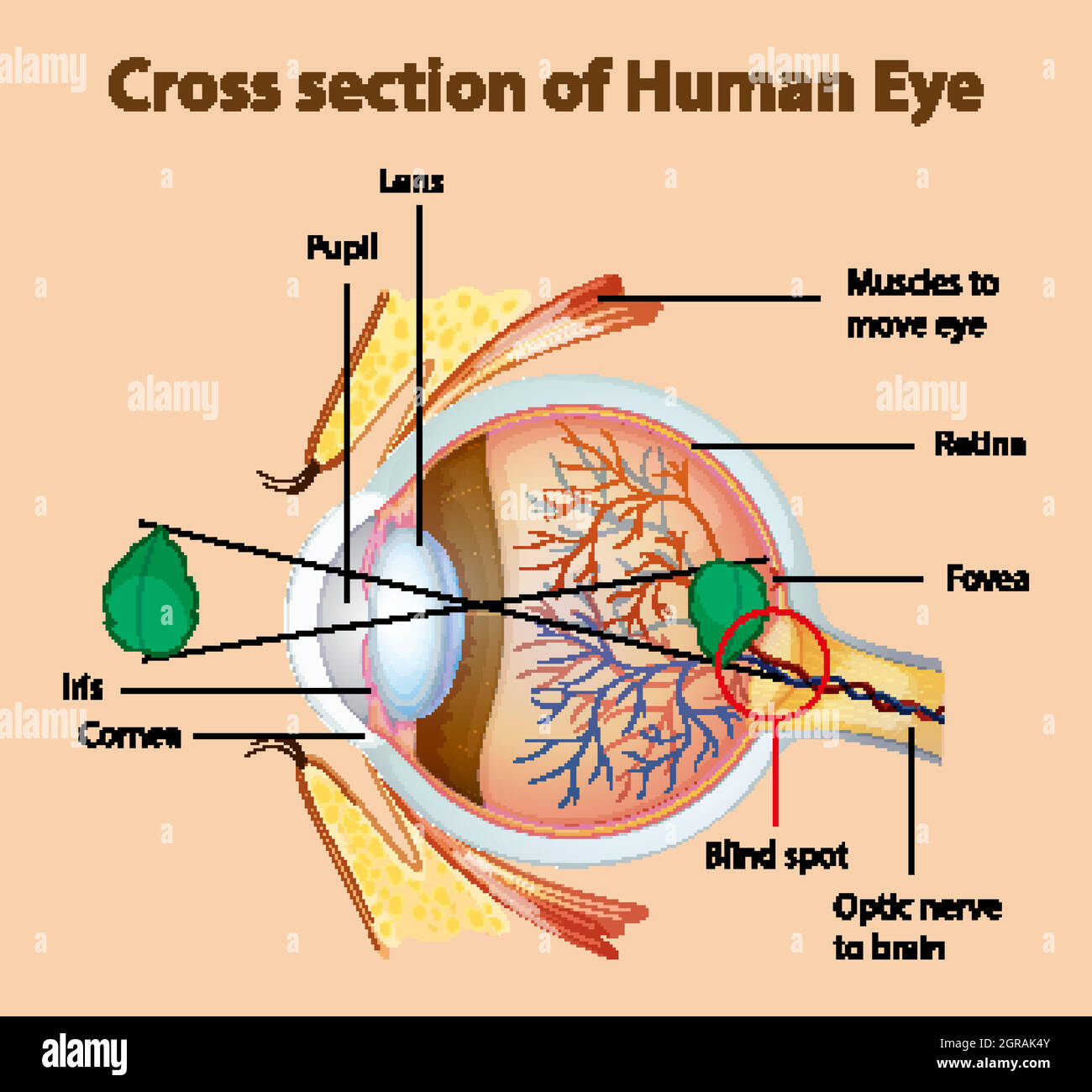
35 eye cross section diagram
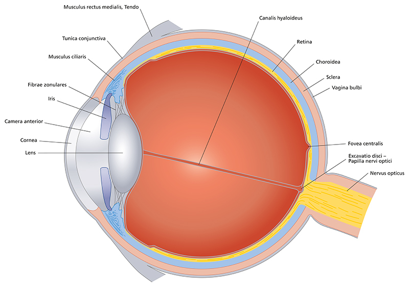
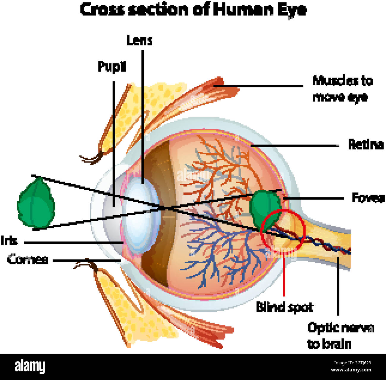
Download this Free Vector about Diagram showing cross section of human eye, and discover more than 18 Million Professional Graphic Resources on Freepik. #freepik #Vector #Education #Character #Chart Eye Cross-section. When light strikes the eye, the first part it reaches is the cornea, a dome positioned over the center of the eye. The cornea is clear and refracts, or bends, the light passing ...
Cross section of vitreous anatomy. A, Cross section diagram of the eye with emphasis on the anatomical features of the vitreous. The vitreous is most firmly attached to the retina at the vitreous base, and it also has adhesions at the optic nerve, along vessels, at the fovea, and to the posterior lens capsule.

Eye cross section diagram
If you compare the equations for Q above to the equations for calculating the centroid (discussed in a previous section), you will see that we actually use the first moment of area when calculating the centroidal location with respect to an origin of interest.. The first moment is also used when calculating the value of shear stress at a particular point in the cross section. Eye Cross Section Labeled Diagram. An eye cut saggital labeled diagram. Human eye anatomy. Cartoon simple illustration for medical atlas or educational textbook. Cross-section of an eyes. Human eye anatomy. Abstract polygonal light of human eye anatomy. Business wireframe mesh spheres from flying debris. Eye cross section concept. The cross section represents a portion of Earth’s crust. Letters A, B, C, and D are rock units. Igneous rock B was formed after rock layer D was deposited but before rock layer A was deposited. Using the contact metamorphism symbol shown in the key, draw that symbol in the proper locations on the cross section provided to indicate those
Eye cross section diagram. inner layer of posterior wall of eye (see Retina in Cross Section) contains receptors that convert light energy into signals that brain can interpret. Choroid: vascular layer that nourishes outer retina. can be inflamed in autoimmune ("rheumatologic") disorders. Sclera: collagenous outer layer of wall of eye. Müllers muscle: Sympathetically-innervated muscle extending from levator palpebrae superioris to top of tarsal plate. Elevates eyelid, but not nearly as much as levator does. Impaired in Horner Syndrome, but ptosis is mild because Müller's muscle is a weak elevator. Orbital septum: Collagenous curtain connecting frontal bone and upper lid tarsus. Download scientific diagram | Cross sectional diagram of human eye [1]. from publication: Identification of Diabetic Retinal Exudates in Digital Color Images Using Support Vector Machine ... Human eye cross section anatomy diagram with all parts anatomical structure for medical science education and health care. Image Editor Save Comp. Similar Illustrations See All. Human eye anatomy with cross section of eye diagram illustration;
The cross section represents a portion of Earth’s crust. Letters A, B, C, and D are rock units. Igneous rock B was formed after rock layer D was deposited but before rock layer A was deposited. Using the contact metamorphism symbol shown in the key, draw that symbol in the proper locations on the cross section provided to indicate those Eye Cross Section Labeled Diagram. An eye cut saggital labeled diagram. Human eye anatomy. Cartoon simple illustration for medical atlas or educational textbook. Cross-section of an eyes. Human eye anatomy. Abstract polygonal light of human eye anatomy. Business wireframe mesh spheres from flying debris. Eye cross section concept. If you compare the equations for Q above to the equations for calculating the centroid (discussed in a previous section), you will see that we actually use the first moment of area when calculating the centroidal location with respect to an origin of interest.. The first moment is also used when calculating the value of shear stress at a particular point in the cross section.
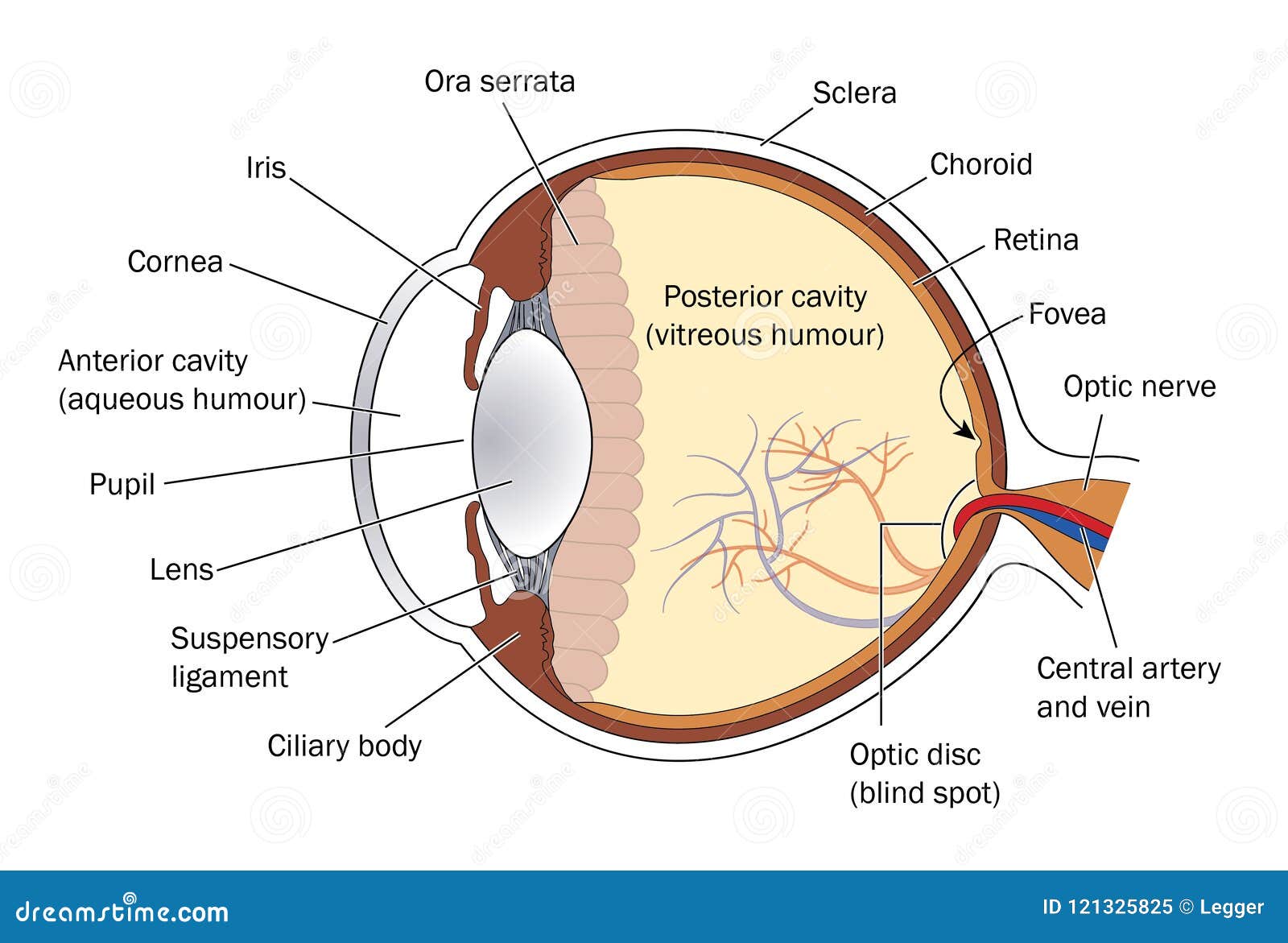
Cross Eye Section Stock Illustrations 469 Cross Eye Section Stock Illustrations Vectors Clipart Dreamstime
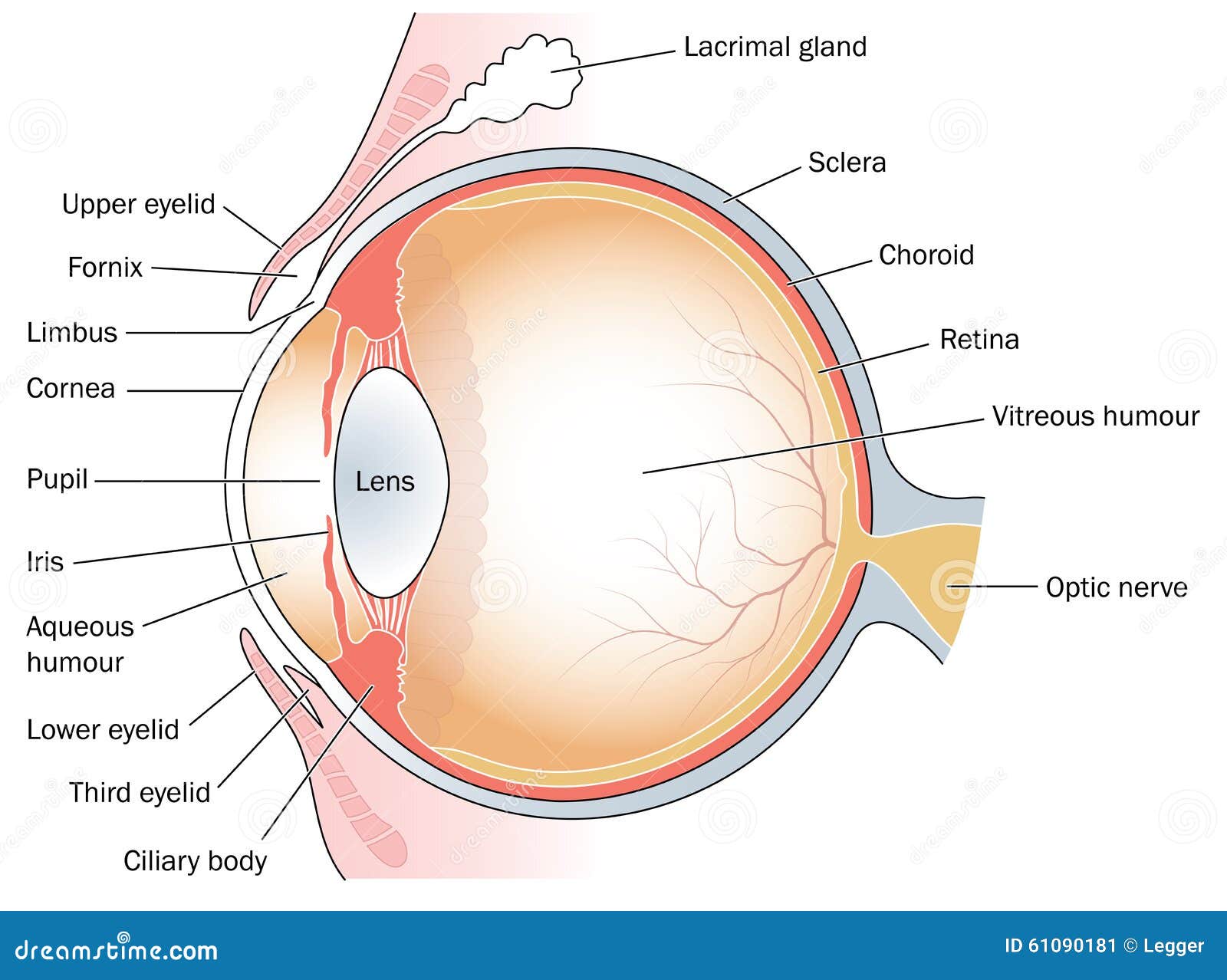
Cross Section Eye Stock Illustrations 469 Cross Section Eye Stock Illustrations Vectors Clipart Dreamstime

Cross Eye Section Stock Illustrations 469 Cross Eye Section Stock Illustrations Vectors Clipart Dreamstime

Eye Anatomy Rod Cells And Cone Cells The Arrangement Of Retinal Cells Is Shown In A Cross Section Vector Diagram For Your Design Educational Biological Science And Medical Use Royalty Free Cliparts

Cross Section Illustration Of Human Eye Stock Photo Picture And Low Budget Royalty Free Image Pic Esy 036249479 Agefotostock











0 Response to "35 eye cross section diagram"
Post a Comment